Figure 1.

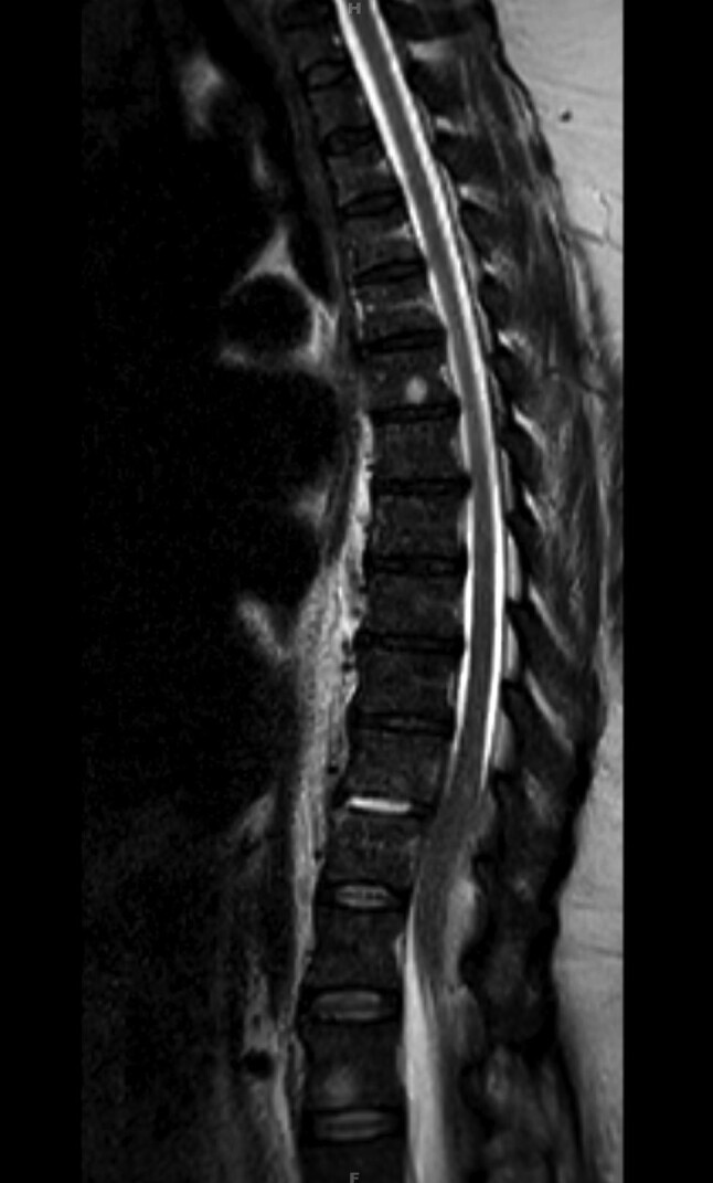
Sagittal view of contrast-enhanced magnetic resonance imaging of thoracic and lumbar spine shows discitis at the T10–T11 disc space level. Abnormal meningeal enhancement is noted at T10–T12, causing some spinal cord compression.

Sagittal view of contrast-enhanced magnetic resonance imaging of thoracic and lumbar spine shows discitis at the T10–T11 disc space level. Abnormal meningeal enhancement is noted at T10–T12, causing some spinal cord compression.