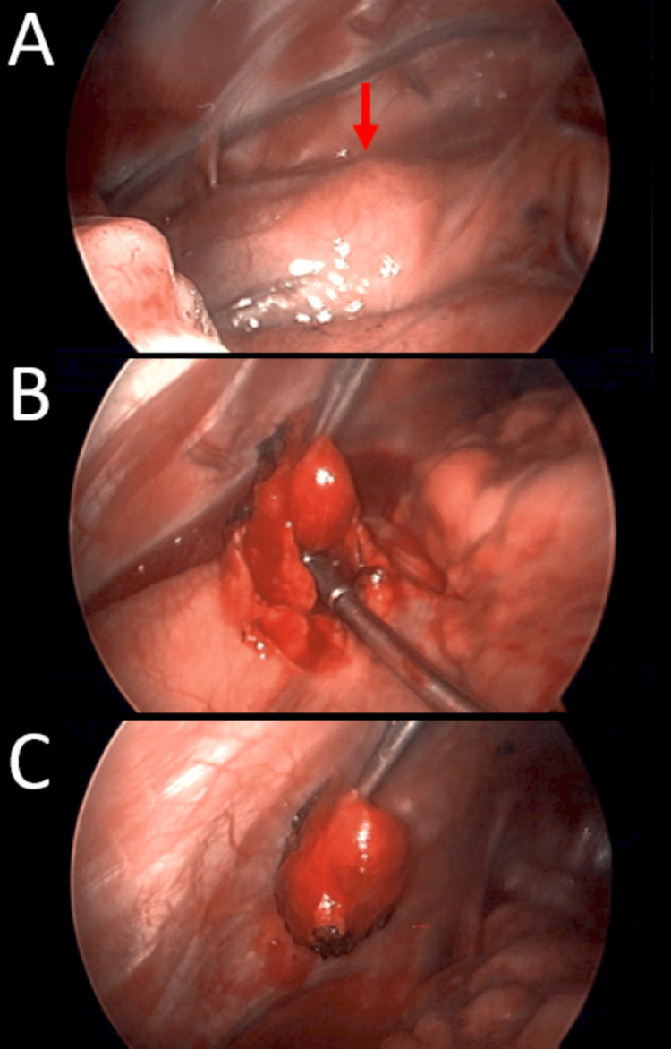
Figure 3. Intraoperative video-assisted thoracoscopic surgery (VATS) demonstrating excision of the causative lesion.
A rounded 1 cm mass was seen in the subpleural space, located below the aortic arch above the heart, and anterior to the phrenic nerve by several millimeters (red arrow, panel A). Electrocautery was lightly applied to the pleura, opening it, and dissection was very carefully performed with thoracic graspers. Gentle elevation with the suction tip was able to remove the lesion with minimal bleeding (panels B and C).

