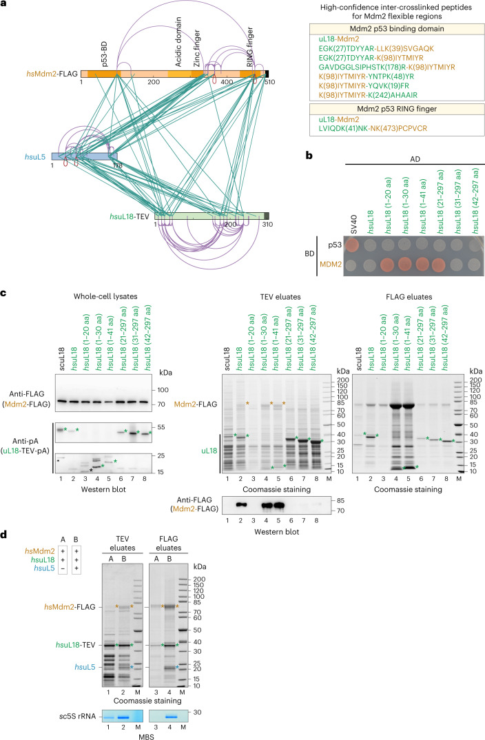
Fig. 6. The human uL18 N-terminal sequence specifically binds to the Mdm2 N domain.
a, Left: XL-MS of the purified Mdm2–5S RNP complex, using DSS-H12. All inter-crosslinks and self-crosslinks are depicted. The xiNET tool45 was used for visualization of the crosslinks and the primary structure of the proteins. The domain organization of Mdm2 is also displayed. Right: manually curated list of the high-confidence inter-crosslinked peptides for the flexible regions of Mdm2 unresolved by cryo-EM. b, Yeast two-hybrid interaction between the indicated hsuL18 constructs and hsMdm2. AD, activation domain; BD, binding domain. The yeast two-hybrid assay was performed twice with a similar outcome. c, Sequence-specific binding of hsuL18 to hsMdm2, analyzed by co-expression and pull-down assays in yeast cells. GAL-induced co-expression of hsuL18 N-terminal constructs (fused at the C terminus to TEV-ProtA) and hsMdm2-FLAG, followed by IgG Sepharose chromatography and TEV cleavage (TEV eluates). Total lysates (left) were analyzed by western blotting for hsuL18-TEV-ProtA and Mdm2-FLAG using anti-ProtA and anti-FLAG antibodies, respectively. The TEV eluates were further affinity purified on FLAG beads to enrich for Mdm2-FLAG. Both TEV (middle) and FLAG (right) elutes were analyzed by SDS–PAGE. Mdm2 and uL18 bands are indicated by orange and green asterisks, respectively. The FLAG-labeled Mdm2 bands in lanes 2, 4 and 5 (orange asterisks) of the Coomassie-stained gel (TEV eluates) were also verified by mass spectrometry. This co-immunoprecipitation assay was performed twice with similar outcomes. d, Cooperative binding of hsuL5 and hsuL18 to Mdm2, analyzed by co-expression and pull-down assays in yeast cells. Sample A corresponds to the co-expression of hsuL18 and hsMdm2, whereas in sample B, hsuL5 was added to the in vivo co-expression system. Tandem affinity purifications from the cell lysates were performed by pulling down hsuL18-TEV-ProtA (TEV eluates) in the first step and hsMdm2-FLAG (FLAG eluates) in the second step. Eluates were analyzed by SDS–PAGE and Coomassie staining or methylene blue staining. Mdm2, uL18 and uL5 bands are indicated by orange, green and blue asterisks, respectively. This co-immunoprecipitation assay was performed twice with similar outcomes.

