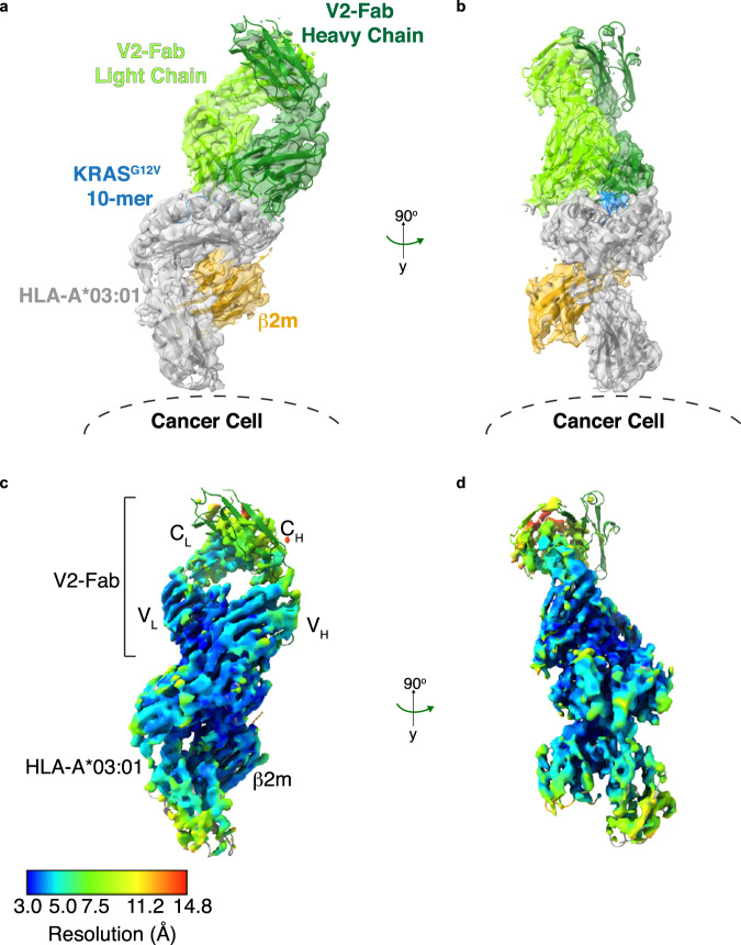
Fig. 2. Structure of V2-Fab/KRASG12V-HLA-A*03:01.
a The final rebuilt model of V2-Fab/KRASG12V-HLA-A*03:01 (PDB ID 7STF), with the cryo-EM map superimposed and in a transparent view. The structure and map are colored according to Fig. 1e. To aid the conceptual visualization, the location of the HLA-A*03:01 relative to the surface of the cancer cell is highlighted. b Structure and density map of V2-Fab/KRASG12V-HLA-A*03:01 at 90° to the view shown in (a). c Local resolution map of V2-Fab/KRASG12V-HLA-A*03:01 in the same orientation as (a). Resolution map was calculated in MonoRes and depicted in ChimeraX. d Local resolution map of V2-Fab/KRASG12V-HLA-A*03:01 at 90° to that shown in (c).

