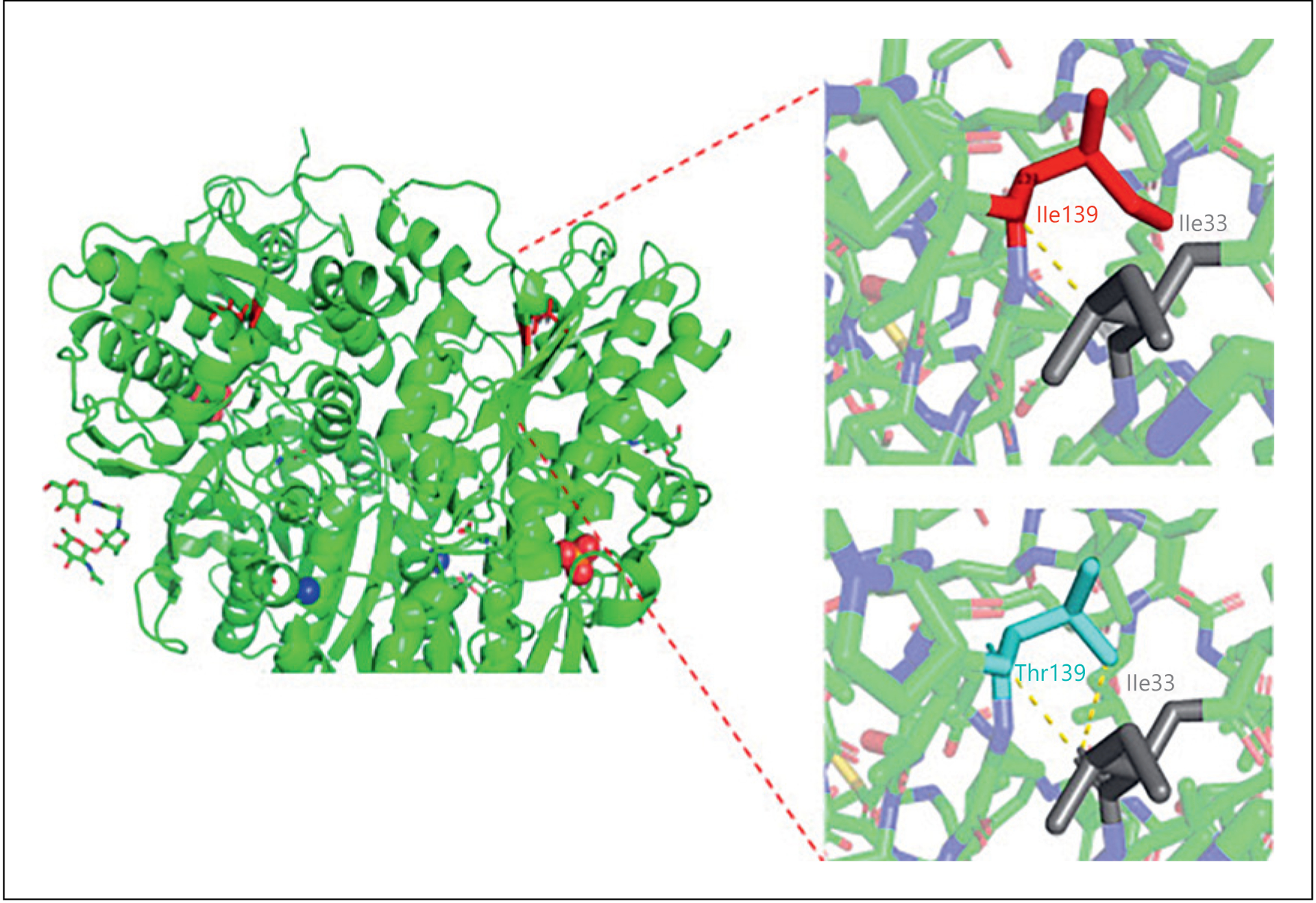
Fig. 2.

Ribbon diagram of the CASR structure (left) and localization of the mutation within the ligand-binding domain of the receptor in the extracellular domain (position 139 in red). Modeling of the wild-type Ile139 (Upper right) and mutant Thr139 residue (Lower right) using an inactive state CASR structure obtained from high-resolution cryo-EM and Pymol showed the side chain changing from large aliphatic residue to a small neutral one. The introduction of a mutant Thr139 residue (pale blue) is predicted to form an additional hydrogen bond between residue Thr139 and Ile33 (dashed yellow lines).
