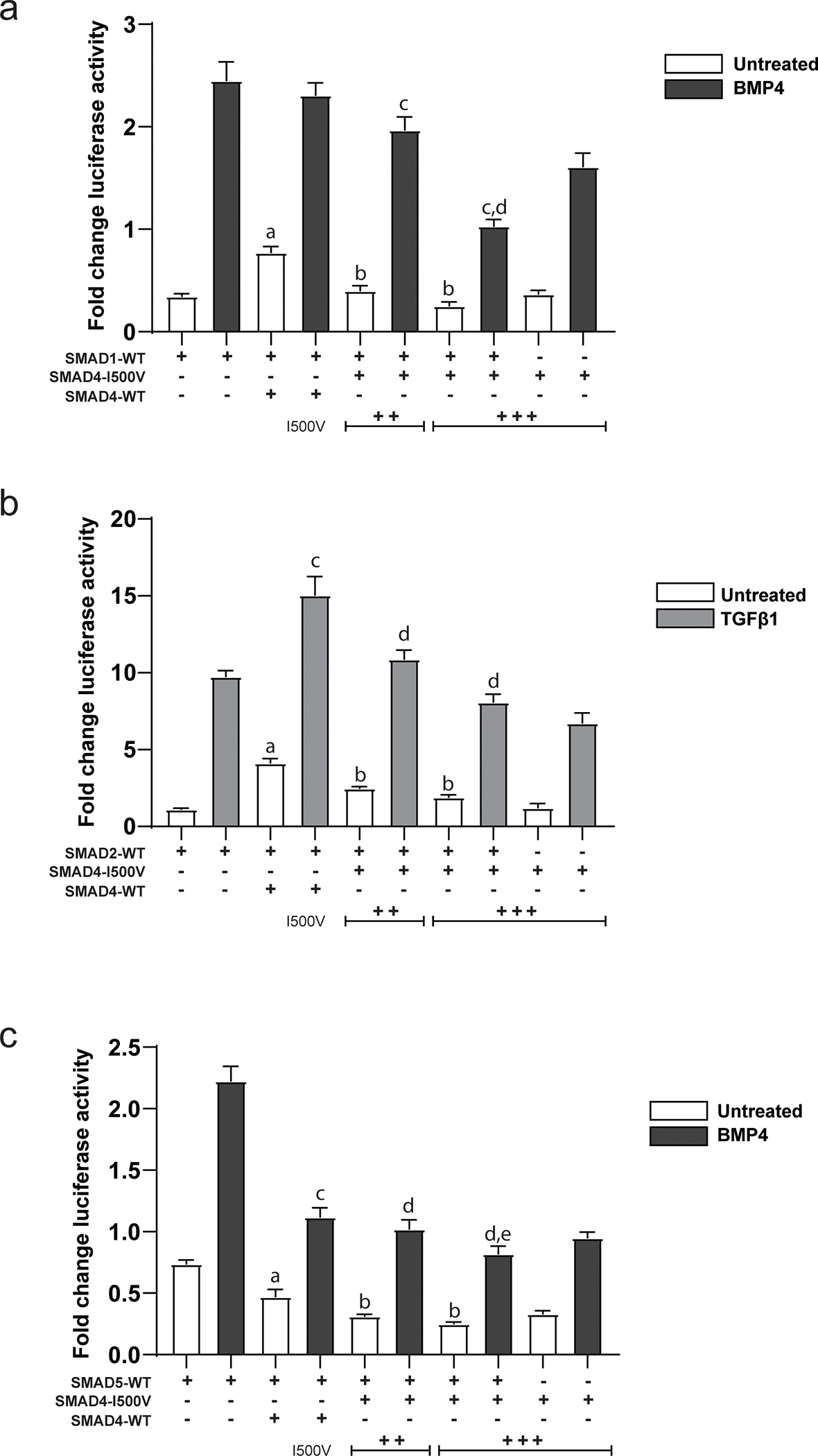
Fig. 2. Dominant-negative activity of SMAD4-I500V inhibits function of R-SMADs.

(A) Action of the I500V variant on SMAD1 activity on the Xvent2-luc reporter in basal and BMP4-treated cells was assessed, a: comparison between SMAD1- and SMAD1/WT-SMAD4- transfected cells in basal conditions, p < 0.0001; b: comparison between SMAD1/WT-SMAD4- and SMAD1/SMAD4-I500V- transfected cells in basal conditions, p < 0.0001; c: comparison between SMAD1/WT-SMAD4- and SMAD1/SMAD4-I500V- transfected cells in BMP4-treated cells, p < 0.0001; d: comparison between SMAD4-I500V- and SMAD1/SMAD4-I500V- transfected cells in BMP4-treated cells, p < 0.0001. (B) Action of the I500V variant on SMAD2 activity on the SBE(4)-luc reporter in basal and TGFβ1-treated cells was assessed, a: comparison between SMAD2- and SMAD2/WT-SMAD4-transfected cells in basal conditions, p < 0.0001; b: comparison between SMAD2/WT-SMAD4- and SMAD2/SMAD4-I500V-transfected cells in basal conditions, p < 0.0001; c: comparison between SMAD2- and SMAD2/WT-SMAD4- transfected cells in TGFβ1-treated conditions, p < 0.0001; d: comparison between SMAD2/WT-SMAD4- and SMAD2/ SMAD4-I500V-transfected cells in TGFβ1-treated conditions, p < 0.0001. (C) Action of the I500V variant on SMAD5 activity on the Xvent2-luc reporter in basal and BMP4-treated cells was assessed, a: comparison between SMAD5- and SMAD5/WT-SMAD4- transfected cells in basal conditions, p < 0.0001; b: comparison between SMAD5/WT-SMAD4- and SMAD5/SMAD4-I500V-transfected cells in basal conditions, p < 0.0001; c: comparison between SMAD5- and SMAD5/WT-SMAD4- transfected cells in BMP4-treated conditions, p < 0.0001; d: comparison between SMAD5/WT-SMAD4- and SMAD5/SMAD4-I500V- transfected cells in BMP4-treated conditions, p < 0.05, 0.0001; e: comparison between SMAD5/SMAD4-I500V- and SMAD4-I500V- transfected cells in BMP4-treated cells, p < 0.001. Luciferase activity was normalized to vector transfected cells and analyzed by one-way ANOVA, presented as mean ± SD, n = 6–9.
