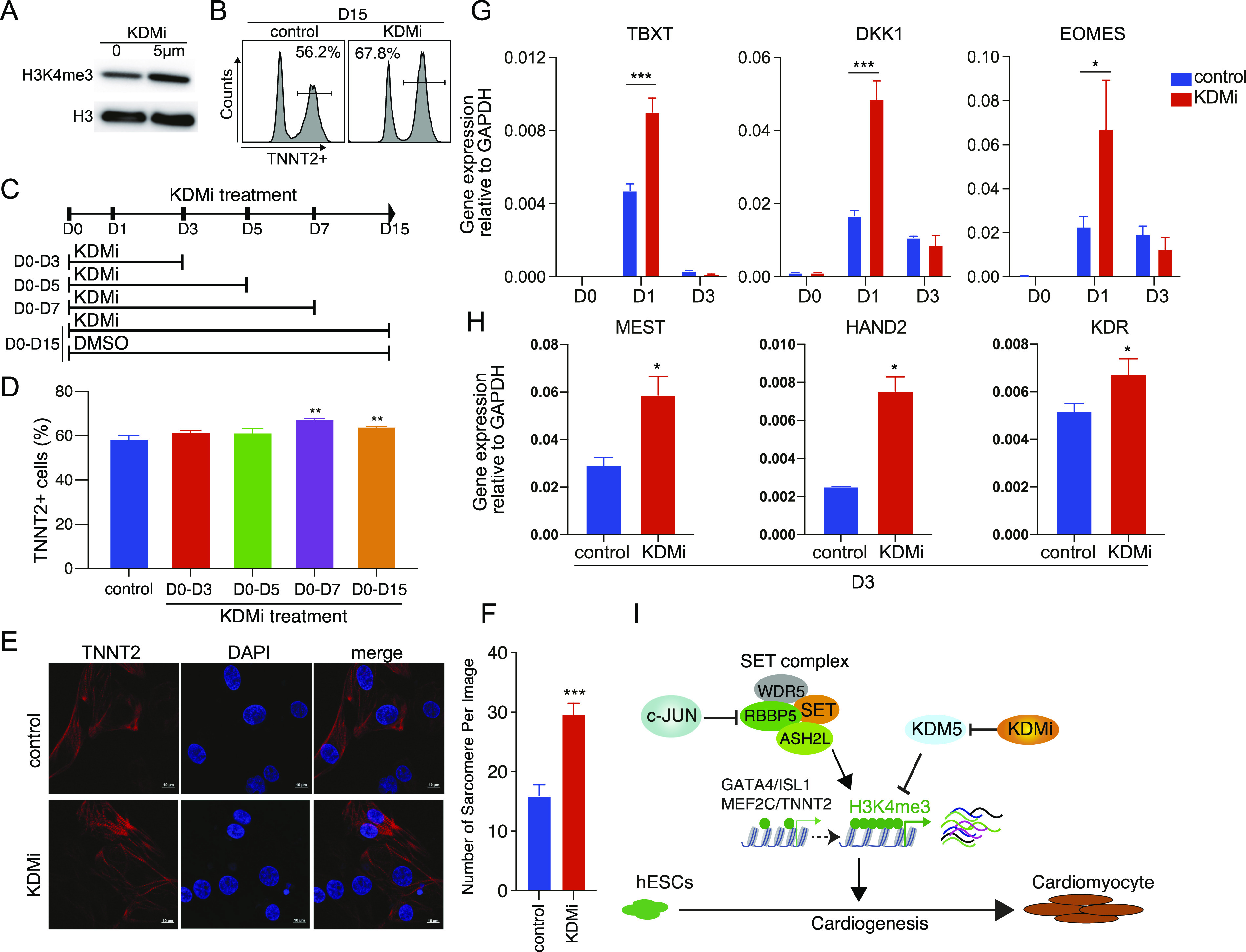
Figure 5. KDM inhibitor promotes hESC differentiation into cardiomyocytes.
(A) Western blot showing the H3K4me3 protein level in hESC treated with 0 μm and 5 μm KDMi. (B) Flow cytometry analysis of TNNT2 expression in cardiomyocytes differentiated from hESC. Percent of cells TNNT2+ is indicated for each condition, control: induced cardiomyocytes from WT hESC with DMSO, KDMi: induce cardiomyocytes from hESC with DMSO-dissolved KDM inhibitor (CPI-455, 5 μm). KDMi was added into the culture medium from D0 to D15. (C) Schematic of the time window of KDMi treatment. (C, D) Flow cytometry quantification of cardiomyocyte differentiation efficiency, based on the percent of TNNT2+ cells in different groups in (C). Data were from eight biological replicates in three independent experiments and are shown as the mean ± SEM. ** P-value < 0.01, two-way ANOVA with Sidak correction between the control and KDMi groups. (E) Immunostaining showing the level of TNNT2 in cardiomyocytes that generated with DMSO or 7-D KDMi treatement (D0–D7). Scale bar, 10 μm. (F) Counts of the number of sarcomeres in each image. Data were from seven to nine replicates and are shown as the mean ± SEM. *** P-value < 0.001, two-way ANOVA with Sidak correction between the control and KDMi groups. (G) Relative expression of the indicated mesoderm marker genes during the differentiation of hESC into mesoderm cells with or without KDMi. Data were from six biological replicates in three independent experiments and are shown as the mean ± SEM. *** P-value < 0.001, * P-value < 0.05, two-way ANOVA with Sidak correction between the control and KDMi groups. (H) Relative expression of the indicated cardiac mesoderm marker genes during differentiation of hESC into cardiac mesoderm cells with or without KDMi. Data were from six biological replicates in three independent experiments and are shown as the mean ± SEM. * P-value < 0.05, Unpaired t test between the control and KDMi groups. (I) Schematic for the role of c-JUN in modulating chromatin accessibility and H3K4me3 modification during hESC to cardiomyocyte differentiation.
Source data are available for this figure.

