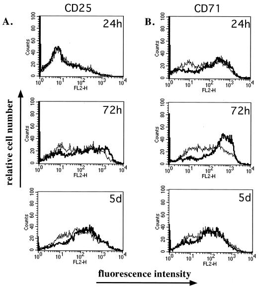
FIG. 3.
Activation markers CD25 and CD71 are expressed on MV-Ed-infected cells following stimulation. Mock-infected (bold curve) or MV-Ed-infected (fine curve) T cells were cultured with PHA and IL-2. At various times, cells were surface labeled with antibodies specific to CD25 or to CD71 followed by a secondary phycoerythrin-conjugated immunoglobulin. Analysis was done by flow cytometry.

