
Fig. 3.
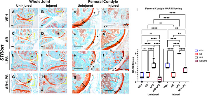
Characterization of posttraumatic osteoarthritis (PTOA) new joint phenotype of STR/ort mice exposed to antibiotics (AB),# lipopolysaccharides (LPS), and a combination (AB+LPS) compared with vehicle (VEH) showing cartilage in red while the bone in bone. (A) VEH uninjured contralateral shows intact morphology with reduced staining in the growth plate. (B, b) VEH injured shows cartilage degradation and tibial degeneration. (C) AB uninjured contralateral shows normal morphology with a lack of growth plate staining. (D, d) AB injured shows thicker femoral condyle cartilage than VEH injured. (E) LPS uninjured showing intact morphology and strong staining of the growth plate. (F, f) Injured LPS showing lack of articular cartilage staining tibial degeneration. (G) AB+LPS showing decrease staining of the femoral condyle but intact morphology. (H, h) AB+LPS injured with thicker cartilage and showing tibial degeneration. (I) Osteoarthritis Research Society International (OARSI) scoring showing significant differences between injury and treatment type (*p < 0.05, **p < 0.01, ***p < 0.001, ****p < 0.00001). # STR/ort AB data in this figure are from the same cohort and same as data presented in Fig. 1; they are compared in different contexts. Scale bars = 200 μm.