Fig. 5.

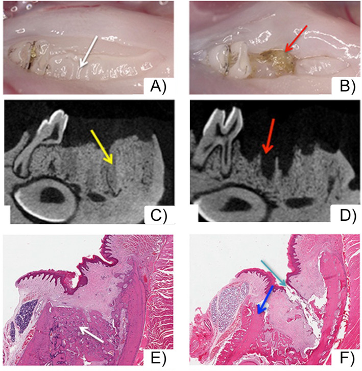
Extraction of healthy teeth or teeth with periapical disease in zoledronate (ZOL)‐treated animals. (A) Clinically extraction sockets of healthy teeth healed with normal mucosa (white arrows). (B) In contrast, extraction sockets of teeth with periapical disease showed areas of bone exposure (red arrows). (C) μCT assessment demonstrated extraction sockets of healthy teeth filled with woven bone (yellow arrow). Socket outlines are clearly demarcated. (D) Extraction sockets of teeth with periapical disease lack bone formation and appear empty, with socket outlines clearly defined (red arrows). (E) Histologic assessment demonstrates woven bone formation in the extraction socket of healthy teeth (white arrow). The margins of the extraction socket and the woven bone are clearly outlined. (F) In contrast, sockets of extracted teeth with periapical disease are void of bone formation, with the socket outlines visible and presence of osteonecrosis (blue arrows), debris, and bone exposure (aqua arrow). (Figure modified from Hadaya and colleagues( 90 )).
