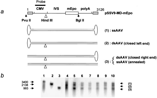
FIG. 3.
Southern blot analysis of the vector DNA after alkaline gel electrophoresis. (a) Positions of restriction sites and probe (CMV promoter) in pSSV9-MD-mEpo. The different species expected to be detected in the analysis are represented below. Three different fragments are expected upon HindIII digestion of these forms: (1) undigested ssDNA, (2) fragment from closed-left-end ds species (ds forms resulting from the synthesis of positive strand), and (3) fragment either from reannealed ds species or from closed-right-end species (ds forms resulting from synthesis of negative strand). IVS, second intron from the human β-globin gene. (b) Southern blot following alkaline gel electrophoresis of HindIII-digested total muscle DNA (4 μg) from animals sacrificed on day 1 (lanes 5 and 6 correspond to two different DNA samples from the same injected muscle split prior to the extraction process, and lane 7 corresponds to a second animal), day 2 (one animal, lane 8), and day 3 (two animals, lanes 9 and 10). Lane 4 contains DNA from 293 cells coinfected with AAV-MD-mEpo and a helper adenovirus as a control for second-strand synthesis. Lanes 1 to 3 contain the following size markers: purified DNA from rAAV green fluorescent protein Neo particles (lane 1), PvuII/BglII fragment from pSSV9-MD-mEpo (lane 2), and PvuII/HindIII fragment of pSSV9-MD-mEpo (lane 3). Sizes in bases are indicated on the left.

