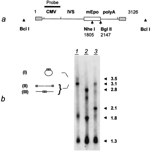
FIG. 4.
Southern blot analysis of rAAV DNA species in an animal sacrificed 1 day after injection. (a) Positions of restriction sites and probe (CMV promoter) in MD-AAV-mEpo (see legend to Fig. 1a). IVS, second intron from the human β-globin gene. (b) Four micrograms of total DNA was digested by BclI (lane 1), BglII (lane 3), or both BglII and NheI (lane 2). The diagram on the left indicates the deduced structure of the 3.5- and 3.1-kb DNA species: circular monomeric form (I), ds monomeric form (linear) (II), or linearized circular form and/or digested head-to-tail concatemer (III). The shaded boxes indicate the ITRs.

