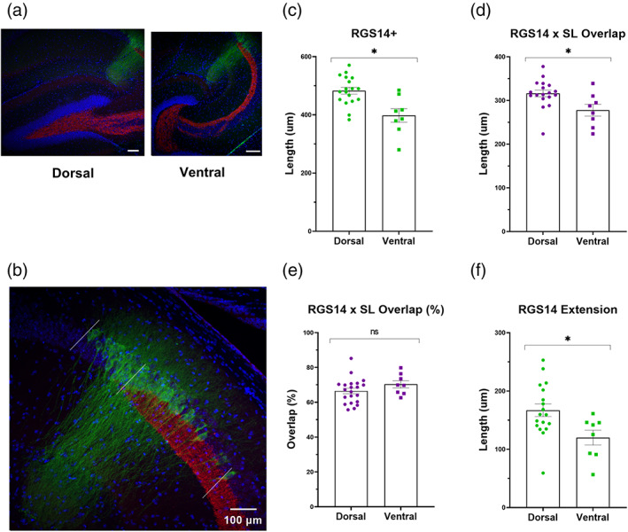
FIGURE 3.

RGS14 expression patterns in relation to Znt3+ mossy fibers (MFs) differ along the dorsal–ventral axis of the hippocampus. (a) Slice position along the dorsal–ventral axis was confirmed by the shape of the dentate gyrus in 10× confocal images as illustrated in the left (dorsal, “V‐shape”) and right (ventral, “C‐shape”) panels. Scale bars = 200 μm (b) 20× confocal image comprising the extent of RGS14 labeling relative to Znt3+ staining. The length between the proximal and distal extent of RGS14+ immunohistochemical staining (outside white lines) was subdivided based on proximity to the distal end of the Znt3+ stratum lucidum (center white line). Scale bar = 100 μm (c) The length of RGS14+ expression is significantly larger in dorsal versus ventral slices (483.14 ± 11.70 vs. 397.95 ± 23.18 μm, p = .001, t = 3.66, f = 1.74, n = 18, 8 sections). (d) On average, the length of RGS14+ staining (green, b) that overlaps with labeled mossy fibers in stratum lucidum (red, b) is significantly longer in dorsal versus ventral slices (316.24 ± 7.58 μm in dorsal slices and 277.83 ± 13.49 in ventral, p = .014, t = 2.66, f = 1.41, n = 18, 8 sections, from 8 animals). (e) The percent overlap, relative to total RGS14+ length, is, on average, similar in dorsal and ventral sections (66.45% and 70.34%, p = .19, t = 1.34, 1.48, n = 18, 8 sections, from 8 animals). (f) The length of RGS14+ staining that extends beyond the distal most tip of stratum lucidum (towards CA1) is significantly longer in dorsal hippocampal slices when compared with ventral slices (166.91 ± 10.77 and 120.13 ± 12.77 μm, p = .017, t = 2.55, f = 1.6, n = 18, 8 sections, from 8 animals).
