FIGURE 7.

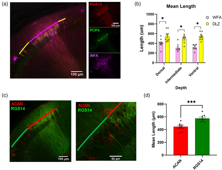
Staining for perineuronal nets does not label the entirety of CA2. (a) Left, Representative image of RGS14, PCP4, and Wisteria Floribunda agglutinin (WFA) immunofluorescence (20× magnification). WFA labeling (magenta) does not completely overlap with the distal‐most and proximal‐most ends of the RGS14+/PCP4+ double‐labeled zone (DLZ; yellow). Right, Confocal images of single stains. (b) Mean length of WFA immunopositivity is significantly shorter than that of the RGS14/PCP4+ DLZ throughout the dorsoventral axis. (dorsal WFA = 405.33 ± 26.10 vs. DLZ = 548.15 ± 33.57 μm p = .004, t = 3.36, f = 1.66, n = 10 sections from 3 animals; intermediate WFA = 308.34 ± 27.83 μm vs. DLZ = 496.42 ± 28.94 μm p = 0004, t = 4.68, f = 1.08, n = 8 sections from 3 animals; ventral WFA = 318.10 ± 18.54 μm vs. DLZ = 541.88 ± 25.06 μm p ≤ .000, t = 7.18, f = 1.83 n = 9 sections, from 3 animals). (c1) Representative image of ACAN and RGS14 immunofluorescence. (c2) Expanded view of inset from (c1). (d) Mean length of Aggrecan immunopositivity is significantly shorter than that of RGS14 (ACAN+ = 575.44 ± 21.10 μm vs. RGS14+ = 446.02 ± 22.76 μm, p = .001, t = 4.17, 1.66, n = 8 dorsal sections, from 3 animals).
