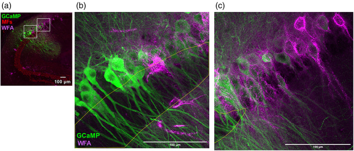
FIGURE 8.

Wisteria Floribunda agglutinin (WFA) most strongly associates with mossy fiber (MF)‐negative CA2 neurons. (a) Reference overview image (40×; 1024 × 1024 pixels) of a horizontal section from dorsal hippocampus showing WFA (magenta) and Znt3 (red) immunopositivity and GCaMP6f‐expressing CA2/CA3 pyramidal neurons. Insets are expanded (2048 × 2048 pixels) in (b) and (c). (b,c) Expanded views from (a) showing WFA‐immunopositive perineuronal nets surrounding GCaMP‐expressing CA2 pyramidal neurons, with Znt3‐immunopositive stratum lucidum outlined in yellow. Intensity of WFA staining is strongest in the MF‐negative CA2 (c) and markedly reduced towards the CA3/CA2 border (b). All scale bars = 100 μm.
