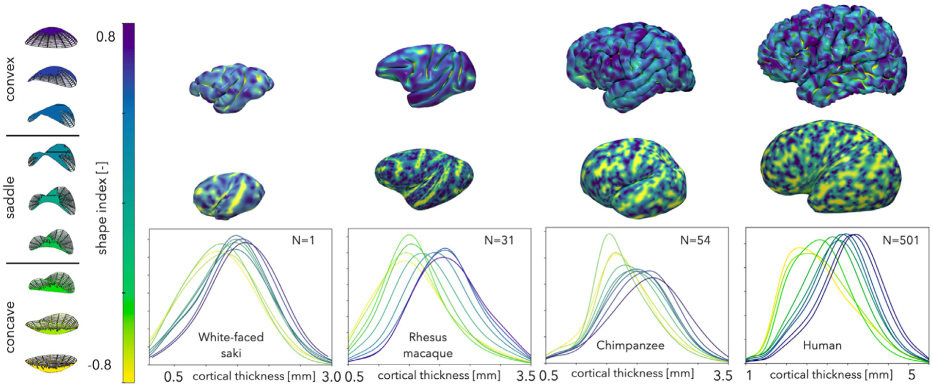
Fig. 5.

Correlation of cortical thickness with cortical geometry. Top: shape index is overlaid onto the pial surface of a representative small (white-faced saki), medium (rhesus macaque), large (chimpanzee), and x-large (human) brain. Middle: Inflated pial surfaces with shape index overlaid. Bottom: Cortical thickness kernel density distribution profiles with respect to local shape, aggregated for N = 1 white faced saki, N = 31 rhesus macaques, N = 54 chimpanzees, and N = 501 humans, are shown. Cortical thickness decreases consistently from convex to saddle to concave shape.
