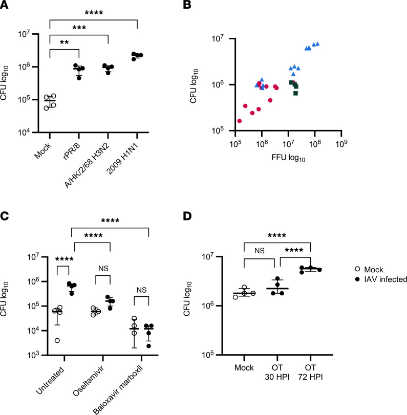
Figure 1. IAV replication increases Spn in the airway.
(A and B) HBECs were infected with 150,000 PFU of IAV for 72 hours, followed by infection with 1,000 CFU of Spn (n = 4–16). (C) HBECs were infected with 100,000 PFU of IAV (pH1N1) and treated basally with 1 mM oseltamivir or 100 nM baloxavir marboxil for 72 hours, followed by infection with 1,000 CFU of Spn (n = 3–4). (D) HBECs were infected with 100,000 PFU of IAV (pH1N1) and then treated basally with 1 mM oseltamivir either 30 or 72 hours after IAV infection. After 72 hours of IAV infection, HBECs were apically infected with 1,000 CFU of Spn. Spn was quantified by vertical plating of apical washes collected after 6 hours of Spn infection (n = 4). All panels except C were analyzed by 1-way ANOVA with Dunnett’s (A) or Tukey’s (C and D) multiple-comparison test. **P < 0.01; ***P < 0.001; ****P < 0.0001. Brackets indicate median and interquartile range. Panel C was analyzed by Pearson’s correlation: r = 0.9348, 95% CI = 0.8645 to 0.9692, P < 0.0001. Open circles indicate mock IAV infection; closed circles indicate IAV infection. In panel B, magenta circles indicate rPR/8, green squares indicate H3N2, and blue triangles indicate pH1N1.

