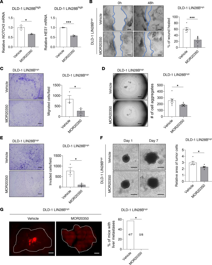
Figure 7. Pharmacologic inhibition of NOTCH3 reduces LIN28B-induced liver metastasis.
(A) qRT-PCR analysis of NOTCH3 and HES1 mRNA in DLD-1 LIN28Bhi cells treated with vehicle or 10 μg/mL MOR20350 for 48 hours. (B) Representative images and quantification of the wound closure scratch assay performed using DLD-1 LIN28Bhi cells treated with vehicle or MOR20350. Scale bar = 200 μm. Area of the wound was measured by using ImageJ. (C) Representative images and quantification of the 3D aggregation assay performed using DLD-1 LIN28Bhi cells treated with the vehicle or MOR20350. The number of cell aggregates was counted using Keyence BZ-X810. (D) Representative images and quantification of 2D invasion assay. Cells that have invaded through the 8 μm pore and the ECM were counted using Keyence BZ-X810. (E) Representative images and quantification of 2D migration assay. Cells that have migrated through the 8 μm pore were counted using Keyence BZ-X810. (F) Representative images and quantification of the 3D invasion assay performed using DLD-1 LIN28Bhi cells treated with the vehicle or MOR20350. Scale bar = 500 μm. (G) Representative images of RFP expressed by DLD-1 LIN28Bhi tumors in the liver. Images were taken using Keyence BZ-X810. Scale bar = 500 μm. Data in graphs A–F are represented as means ± SEM and were analyzed by 1-way ANOVA followed by Tukey’s multiple-comparison test. Data in G were analyzed by Fisher’s exact test. *P < 0.05, ***P < 0.001.

