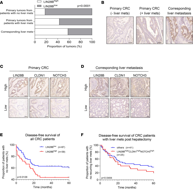
Figure 8. LIN28B/CLDN1/NOTCH3 axis positively correlates with metastatic progression of human colorectal tumors.
(A) Primary colonic tumors and liver metastases were collected from patients with CRC. Upon IHC staining, tumors were quantified based on their high or low expression. Data expressed as a proportion of all tumors (%). (B) Representative images of tumors stained with an anti-LIN28B antibody. Scale bar = 100 μm. (C) Representative images of primary colorectal tumors stained with LIN28B, CLDN1, and NOTCH3. Scale bar = 100 μm. (D) Representative images of corresponding liver metastases stained with LIN28B, CLDN1, and NOTCH3. Scale bar = 100 μm. (E) Graph depicting disease-free survival of all patients with CRC. For all patients, frequency with which they developed liver metastases was tracked over 5 years. Data expressed as the proportion of patients with CRC who did not have metastatic liver tumor (y axis) at the time of analyses (x axis). (F) Graph depicting disease-free survival of patients with CRC who had undergone hepatectomy due to liver metastases. For all patients, frequency with which they developed a new liver tumor was tracked over 5 years. Data expressed as the proportion of patients with CRC who did not develop another metastatic liver tumor (y axis) at the time of analyses (x axis). “Others” group (blue line) includes patients with tumors that do not have high expressions of LIN28B, CLDN1, and NOTCH3. Data in graph in A were analyzed by χ2 test. Data in E and F were analyzed by log-rank test.

