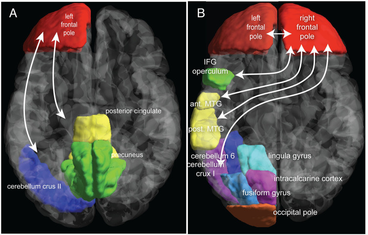
Fig 8. Illustration of the location of regions with increased functional connectivity to frontal poles following use of bright light therapy.
A. Percent bright light therapy use and enhanced functional connectivity between left frontal pole and two clusters: one containing the precuneus and posterior cingulate cortex and the other the cerebellum crus2. B. Percent bright light therapy use and enhanced functional connectivity between right frontal pole and five clusters. The largest included cerebellum crus1, cerebellum 6, occipital pole, lingula gyrus, fusiform gyrus and intracalcarine cortex. The other clusters contained the inferior frontal gyrus (IFG) operculum, anterior and posterior middle temporal gyri (MTG) and right and left frontal poles.

