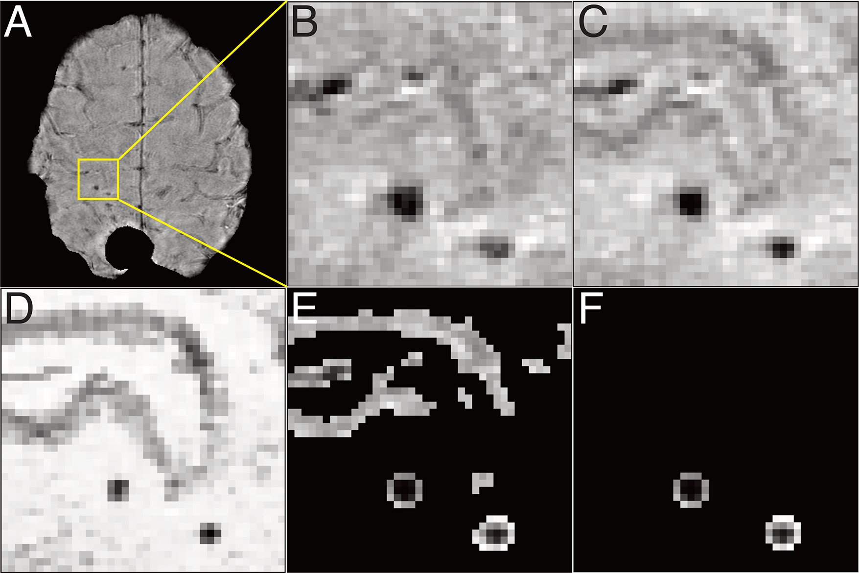
Figure 1. Image processing.

A. whole magnitude image, after removing artifacts due to a prior resection cavity and tumor for input in training. B. zoomed magnitude image, C. zoomed SWI, D. zoomed difference image of SWI and magnitude image, D. zoomed vessel and CMB-masked SWI, E. zoomed CMB-masked SWI.
