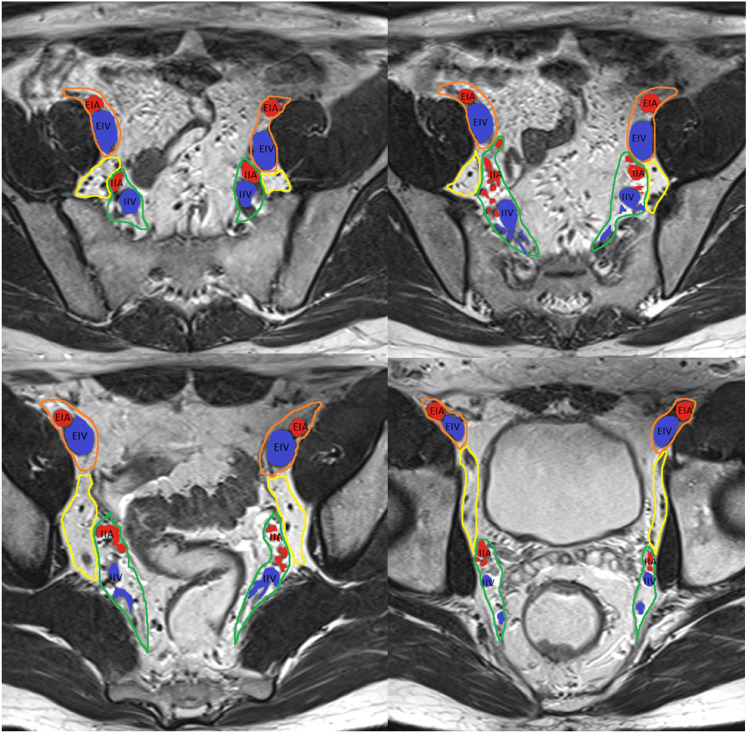
Fig. 4.
Pelvis side wall node anatomy, from upper to lower pelvis. External iliac arteries (EIA, red), external iliac vein (EIV, blue), internal iliac arteries (IIA, red), and internal iliac veins (IIV, blue), and their relationship with the external iliac lymph node region (orange), internal iliac lymph node region (green), and obturator lymph node region (yellow) are depicted. At the level of the obturator muscle, nodes medial to the internal iliac artery are internal iliac lymph nodes (region outlined in green). Lymph nodes lateral to the internal iliac artery within the yellow boundary are obturator lymph nodes [33]

