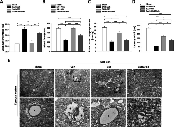
Fig. 2.
Effects of DPSC-CM on brain edema and microcirculation at 24 h post-SAH. A DPSC-CM administration significantly reduced the brain water content in the cortex region at 24 h after SAH. B The regional cerebral blood flow and C the partial pressure of oxygen (PbtO2) at the brain surface were significantly higher in the SAH + CM rats than in the SAH + Veh rats. However, the administration of IGF-1 neutralizing antibodies moderately blunted the DPSC-CM-mediated effects on the two parameters. D DPSC-CM significantly improved the latency to fall at 7 days after SAH induction, as measured by the Rotarod test. E The arrow points to microvessels in representative electron micrographs (bar = 5 μm) from the four groups. L marks the lumen of a microvessel and asterisks mark the end-feet of astrocytes in the higher-magnification images of the lower panel (bar = 1 μm). The swollen end-feet (*) remarkably compressed the microvessels in the SAH + Veh and SAH + CMIGFab groups, whereas DPSC-CM administration attenuated astrocyte swelling. Data are expressed as means ± SEM. *P < 0.05, **P < 0.01, ***P < 0.001, n = 4–5

