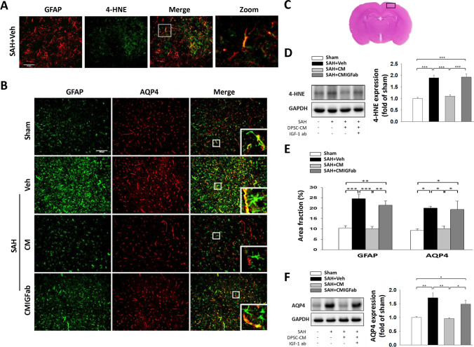
Fig. 3.
DPSC-CM administration reduced the expression of AQP4 and 4-HNE in astrocyte at 24 h post SAH. A Representative immunofluorescence images of 4-HNE (the product of lipid peroxidation; green) and GFAP (a marker for astrocyte; red) labeling in the cerebral cortex region from a SAH animal are shown. Remarkably 4-HNE accumulation was identified in astrocytes after SAH induction (bar = 50 μm). B Representative immunofluorescence images of GFAP and AQP4 labeling in the cerebral cortex region. GFAP immunoreactivity is shown in green, and AQP4 is shown in red (bar = 100 μm). C Representative HE-stained coronal sections from a sham control showing the cortex region to compare the fluorescent signals between the 4 groups of rats, as indicated by the black square boxes. Western blot analysis showed that DPSC-CM administration reduced the expressions of D 4-HNE and F AQP4 in the cortical region at 24 h after SAH. E GFAP and AQP4 expressions were quantified using the area fraction (percentage of GFAP or AQP4 immunoreactivity in the overall field). Data are expressed as means ± SEM. *P < 0.05, **P < 0.01, ***P < 0.001, n = 5

