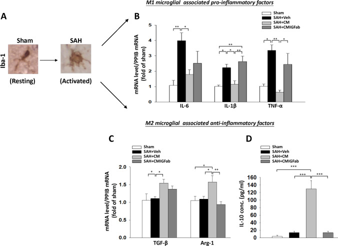
Fig. 4.
Effects of DPSC-CM on pro- and anti-inflammatory microglial (M1-, M2-like) states at 24 h post-SAH. A Representative immunostaining with Iba-1 in the cerebral cortex region and a morphological change from a “resting” form in the sham animal to an “activated” morphology in a SAH animal. B Real-time PCR analysis showed that DPSC-CM administration reduced the mRNA expression of M1 microglial-associated pro‐inflammatory factors, including IL-6, IL-1β, and TNF-α. C SAH induced the mRNA levels of M2 microglial-associated anti‐inflammatory factors, including TGF-β and Arg-1, which were significantly increased in the DPSC-CM group. D ELISA of the protein levels of IL-10 in the four groups showed that DPSC-CM administration significantly increased the IL-10 expression in plasma samples. Data are expressed as means ± SEM. *P < 0.05, **P < 0.01, ***P < 0.001, n = 5

