Fig. 5.
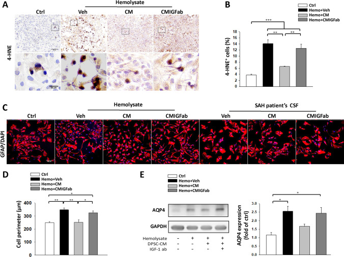
Effects of DPSC-CM on primary astrocytes exposed to hemolysate and SAH-patient CSF for 24 h. A Immunocytochemical staining for 4-HNE (shown in brown; upper panels, bar = 100 μm; lower panels, bar = 20 μm). B There was a significant decrease in the number of 4-HNE-positive cells in the hemolysate + DPSC-CM group. C Immunofluorescence staining for GFAP (shown in red). Representative images showing the effects of exposure of astrocytes to hemolysate or SAH-patient CSF with Veh/DPSC-CM/DPSC-CMIGFab treatment for 24 h, respectively (bar = 100 μm). D Quantitative analysis of GFAP+ cells showed that the astrocyte cell perimeter significantly increased after exposure to hemolysate for 24 h. DPSC-CM treatment significantly decreased the astrocyte cell perimeter. E The level of AQP4 protein in co-cultured astrocytes and microglia was measured by western blot. Hemolysate caused an increase in AQP4 expression, which was suppressed by DPSC-CM treatment. Data are expressed as means ± SEM. *P < 0.05, **P < 0.01, ***P < 0.001, n = 3

