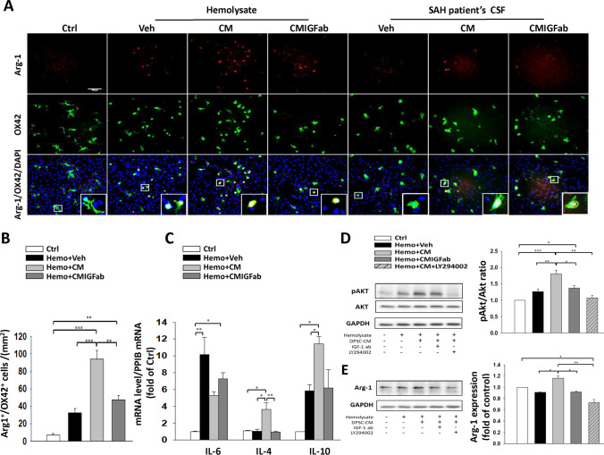
Fig. 6.
Effects of DPSC-CM treatment on microglial M2 polarization in astrocyte/microglia co-cultures exposed to hemolysate or SAH-patient CSF for 24 h. A Representative immunofluorescence images of Arg-1 and OX42 labeling in the astrocyte/microglia co-cultures. OX42 (a marker for activated microglia) immunoreactivity is shown in green, and Arg-1 (M2a microglia marker) is shown in red (bar = 100 μm). B Neutralization of IGF-1 moderately blunted the DPSC-CM-mediated microglia M2 polarization and decreased the number of Arg-1/OX42-positive cells after exposure to hemolysate. C Real-time PCR analysis showed that DPSC-CM markedly inhibited IL-6 and increased the IL-4 and IL-10 mRNA levels after exposure to hemolysate. Western blots and quantification showed that DPSC-CM treatment increased D pAKT (serine-473)/AKT ratio and E the expression of Arg-1 after exposure to hemolysate, which were both reversed by the neutralizing IGF-1 antibody and Ly294002 (AKT-PI3K inhibitor). Data are expressed as means ± SEM. *P < 0.05, **P < 0.01, ***P < 0.001, n = 2–3

