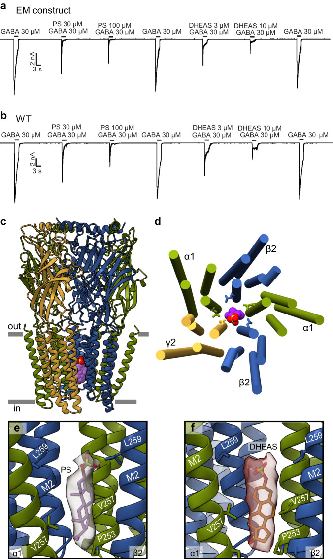
Fig. 4. Sulfated neurosteroids act as pore blockers.
a, b Whole cell patch-clamp electrophysiology recordings comparing the sulfated neurosteroid responses of the EM construct to the full-length WT receptor. c Side view of GABAA receptor – PS complex structure. d View of TMD from extracellular side with PS (colored spheres) bound in the pore. Side chains are shown for 9’Leu residues that form activation gate. e Detailed view of PS in pore site with experimental density shown as semi-transparent surface. f DHEAS in the pore as for PS in (e). In (e) and (f), the γ-subunit is removed for clarity. c–f Subunits are colored as in Fig. 2, with PS in purple and DHEAS in scarlet.

