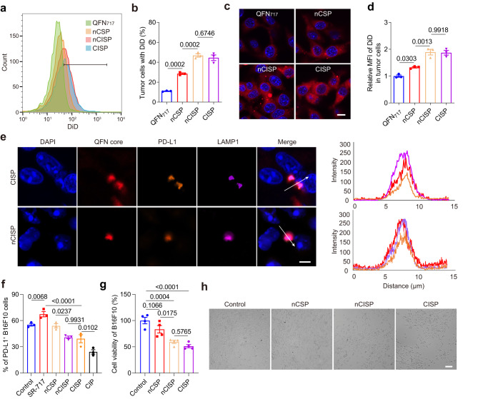
Fig. 2. CISP blocked the elevated PD-L1 in tumor cells induced by STING agonists in vitro.
a, b The uptake of QFN717, nCSP, nCISP, and CISP (marked with DiD) by B16F10 cells detected by flow cytometry (a), and the proportion of cells with DiD (b) (n = 3 independent samples). c, d Representative fluorescence images of B16F10 cells treated with QFN717, nCSP, nCISP, and CISP (marked with DiD, red) (c), and the relative mean fluorescence intensity of DiD in cells (d) (n = 3 independent samples). Scale bar = 10 μm. e Representative images of QFN core, PD-L1, and LAMP1 in B16F10 cells, and the fluorescence intensity on the arrow line (n = 3 independent samples). Scale bar = 5 μm. f Membrane PD-L1 level detected by flow cytometry (n = 3 independent samples). g, h Cell viability of B16F10 detected with CCK-8 kit (g), and representative images of B16F10 cells (h). B16F10 cells were treated with nCSP, nCISP, and CISP, and then incubated with activated CD8+ T cells for 48 h (n = 4 independent samples). Scale bar = 100 μm. Data represent mean ± SEM (b, d, f, g). Statistical significance was determined by a one-way ANOVA test in b, d, f, g and it was two-sided and adjustments were made for multiple comparisons. The experiments for a, b, e–h were repeated three times independently with similar results. Source data are provided as a Source Data file. MFI mean fluorescence intensity, QFN Quercetin-ferrum nanoparticles, QFN717 QFN loaded with SR-717, nCSP normal-Cholesterol cell membrane-coated QFN717, CSP low-cholesterol membrane-coated QFN717, nCISP normal-Cholesterol cell membrane-coated ICB agent and QFN717, CISP low-cholesterol membrane-coated ICB agent and QFN717, CIP low-cholesterol membrane-coated ICB agent and QFN.

