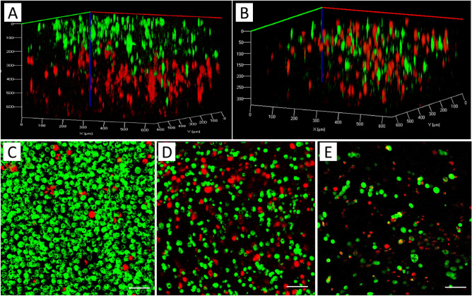
Figure 3.
3D rendered confocal images of MCF7 (stained green) and MDA-MB-231 cells (stained red): (A) Sequential printing of MDA-MB-231 and MCF7, (B) bioprinting of a construct using a bioink with a mixture of MCF7 and MDA-MB-231 cells; 2D culture of both cells mixture with the ratio of 2:1 (MCF7, green:MDA-MB-231, red): (C) Edge of the culture chamber, (D) Middle of the chamber, (E) 3D bioprinting of mixture cells with the ratio of 2:1 (green:red). Scale bars, 50 µm.

