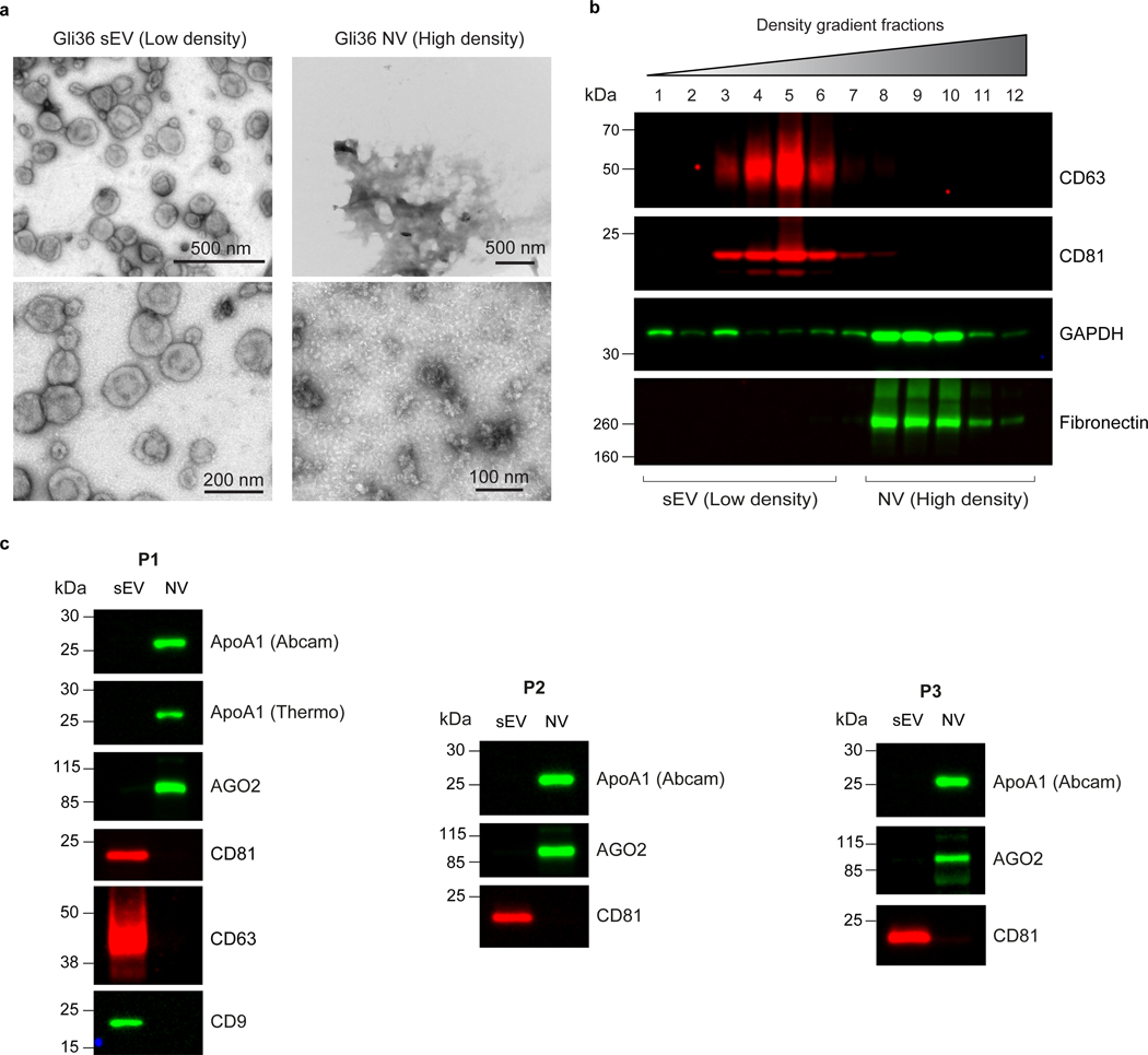
Fig. 6. High-resolution density gradient fractionation separates small extracellular vesicles from non-vesicular components.
a, Negative stain transmission electron microscopy (TEM) of pooled sEV (low density) and NV (high density) fractions derived from Gli36 cells obtained from high-resolution density gradients. The purified sEVs display the expected cup-shaped morphology for EVs while the NV fractions display few distinct structures. b, Density-gradient fractionation of crude small EV pellet (sEV-Ps) derived from DKO-1 cells. After flotation of the sample in high-resolution iodixanol gradients (12 – 36%), equal volumes of each fraction are loaded on SDS-PAGE gels, and membranes were immunoblotted with indicated antibodies. NV, non-vesicular; sEV, small EV. c, High-resolution density gradient fractionation of crude human plasma sEV-Ps to obtain purified sEV and NV fractions. Immunoblots of plasma samples from three normal human individuals (P1–3) following fractionation. ApoA1 (marker for HDL particles) and AGO2 are enriched in the NV fraction while CD81, CD63 and CD9 (markers for sEVs) are enriched in the sEV fraction. The images (panels a-c) are modified from Jeppesen et al.9.

