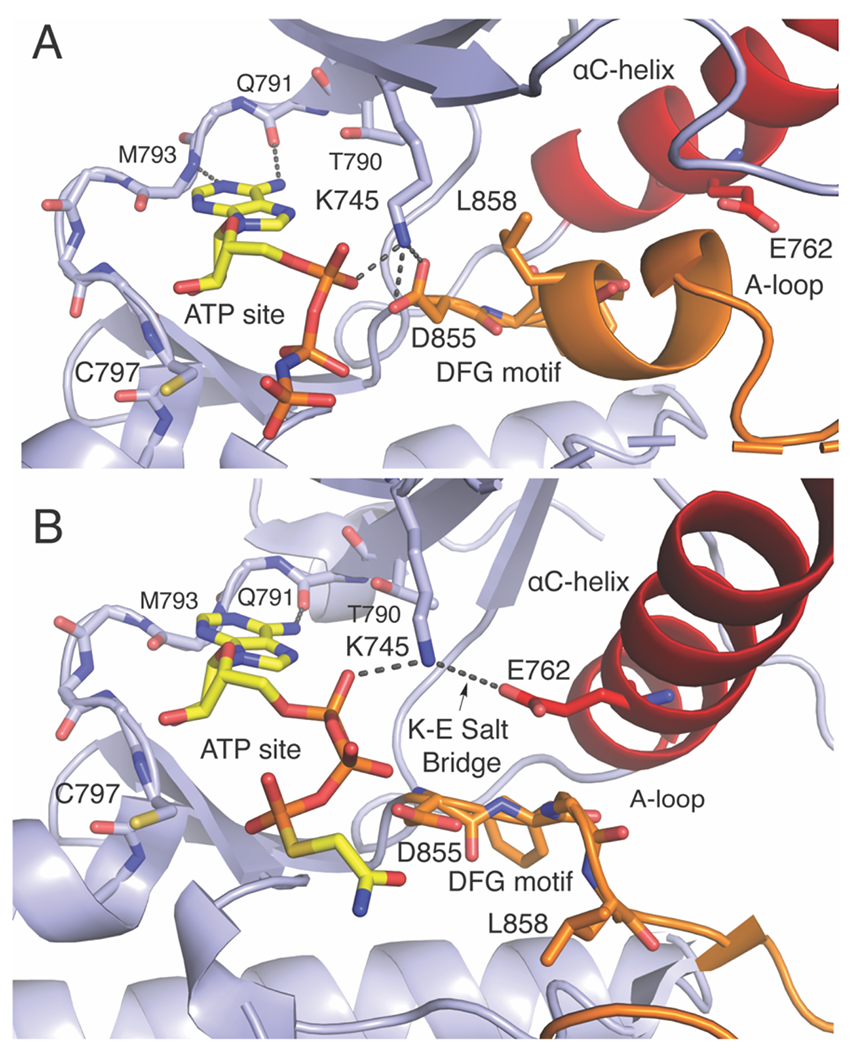
Figure 2. The interplay of residues and binding pockets in the inactive and active EGFR kinase domain.

The EGFR kinase domain in the A) inactive (PDB ID 2GS7, ATP site defined by AMP-PNP is colored yellow) and B) active conformations (PDB ID 2GS6, ATP site defined by an ATP analog-peptide conjugate). Activation loop (A-loop) is colored orange and αC-helix colored red. Some hinge region protein side chains are omitted for clarity
