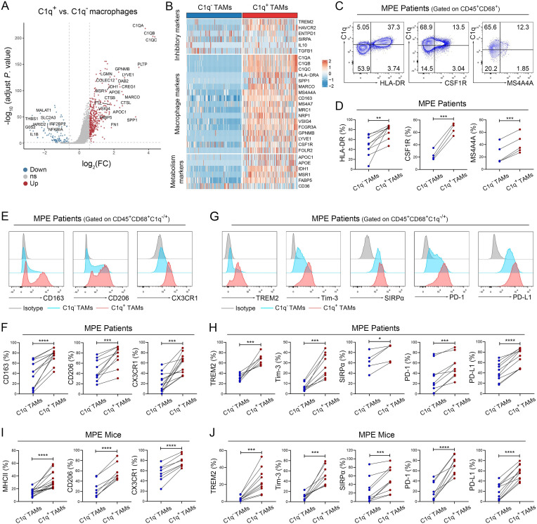
Figure 2.
C1q+ TAMs are characterized by the higher expression levels of immune inhibitory molecules. (A and B) Differentially expressed genes (A, volcano plots) were identified between C1q+ and C1q– macrophages, including macrophage markers, inhibitory markers and metabolism-related genes (B, heatmap). (C and D) Tumor-infiltrating immune cells were isolated from human MPE, and the macrophage markers of C1q+ TAMs were determined by flow cytometry analysis, including the expression levels of HLA-DR, CSF1R and MS4A4A (n=4–9). (E–H) Tumor-infiltrating immune cells were isolated from human MPE; M2-like markers (CD163, CD206, CX3CR1) and inhibitory molecules (TREM2, Tim-3, SIRPα, PD-1, PD-L1) were detected by flow cytometry (n=5–10). (I and J) Tumor-infiltrating immune cells were isolated from mouse MPE; M2-like markers (MHC-II, CD206, CX3CR1; H) and inhibitory molecules (TREM2, Tim-3, SIRPα, PD-1, PD-L1; I) were detected by flow cytometry (n=8–13). Data shown in (C–J) are representative of at least three independent experiments (mean±SD). Statistical analysis was performed using paired two-tailed Student’s t-test (D, F, H–J). *p<0.05, ***p<0.001, ****p<0.0001. CSF1R, colony stimulating factor 1 receptor; CX3CR1, C-X3-C motif chemokine receptor 1; C1q, component 1q; IL, interleukin; HLA, human leukocyte antigen; MHC, major histocompatibility complex; MPE, malignant pleural effusion; PD-1, programmed cell death-1; PD-L1, programmed cell death-ligand 1; SIRPα, signal regulatory protein α; TAM, tumor-associated macrophage; Tim-3, T-cell immunoglobulin-3; TREM2, triggering receptor expressed by myeloid cells-2.

