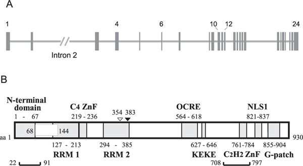
Fig. 1.
Gene and Domain Structure of RBM10. (A) RBM10 gene structure (NCBI Reference Sequence: NG_012548.1). The RBM10 gene contains 24 exons, indicated by vertical closed boxes, with some of these numbered. Horizontal lines between exons indicate introns. Intron 2 is indicated with its relative length shortened by two-sevenths. (B) Schematic domain structure of RBM10. The structure of isoform 1 (930 aa residues) is shown. RBM10 has two RNA-binding domains in the N-terminal region: the RRM1-C4 ZnF di-domain and RRM2 domain. RBM10 isoforms 1 – 4 are determined by the inclusion or exclusion of the region encoded by exon 4 (box at residues 68–144) and of a Val residue at 354 (open triangle) (see also Fig. 3). The brackets at the bottom (residues 22–91 and 709–797) indicate the regions where phosphorylation occurs at multiple sites. K383 (closed triangle) undergoes acetylation/ubiquitylation. Note that the beginning and ending residue-numbers of the domains may not be strictly defined.

