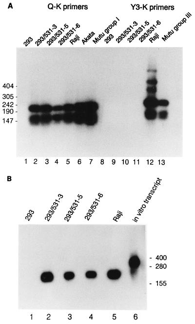
FIG. 5.
Detection of EBNA1 mRNA and EBER1 RNA in infected 293 cells. (A) Spliced EBNA1 transcripts were detected in 2 μg of total RNA from the indicated cell lines by RT-PCR using primers specific for the 3′K (coding) exon and for one of the upstream exons, either Q or Y3 as indicated, to reveal type I or type III latency promoter usage (46), as previously described (26). Amplification products were detected by Southern analysis using labeled internal exon U as a probe. Positions of DNA size markers are indicated (in base pairs) at the left. (B) EBER1 RNA was detected by Northern analysis of 20 μg of total RNA extracted from each cell line. An in vitro transcript (lane 6) includes the EBER1 sequence and additional RNA from the vector, p386 (54). Positions of RNA size markers are shown (in bases) at the right.

