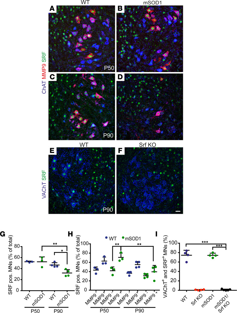
Figure 1. SRF is present in vulnerable MNs.
(A–D) Ventral horns from P50 (A and B) or P90 (C and D) WT (A and C) or mSOD1 (B and D) mice were stained for ChAT (blue), MMP9 (red), and SRF (green). SRF was present in nonvulnerable MMP9– and vulnerable MMP9+ MNs in WT and mSOD1 mice. (E and F) In Srf KO MNs (F), SRF was absent from VAChT+ MNs at P90 in contrast to WT MNs (E). (G) In WT and mSOD1 mice, approximately 50% of MNs were SRF+ at P50. At P90, abundance of SRF+ MNs was decreased in mSOD1 mice. There was no significant change in WT MNs between P50 and P90. (H) In WT mice, SRF was more present in MMP9+ MNs (~60%) compared with MMP9– MNs (~40%). In mSOD1 mice, SRF was significantly more present in vulnerable MMP9+ MNs compared with MMP9– MNs at P50, in contrast to P90. (I) SRF was nearly absent from MNs in Srf-KO and SOD1/Srf-KO mice compared with WT and SOD1 animals at P90. In G–I, each dot represents 1 mouse. (G and H) n = 486 MNs (WT, P50), 377 MNs (mSOD1, P50); 512 MNs (WT, P90), 411 MNs (mSOD1, P90). (I) n = 598 MNs (WT), 558 MNs (Srf KO), 584 MNs (mSOD1), and 650 MNs (mSOD1/Srf KO). Statistical testing was performed by 1-way ANOVA with Tukey corrections. Scale bar: 30 μm (A–F). *P < 0.05, **P < 0.01, ***P < 0.001.

