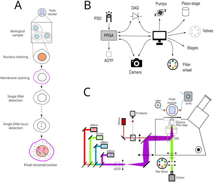
Figure 1.
A. Principle of Hi-M imaging. A single region of interest within the biological sample contains several cells. In cycle 0, a DNA specific dye is used to label the nucleus and other markers. In the following cycles, different probes are injected and imaged. Data from all cycles are combined to reconstruct the final image. B. Overview of hardware devices that are addressed by Qudi-HiM. C. Schematic drawing of a Hi-M microscope. The setup shown here includes 4 excitation lasers (405, 488, 561, 640 nm), an acousto-optic tunable filter, flat and dichroic mirrors, a microscope objective, emission filters, microscope camera, a 785 nm infrared laser and detector, sample holder with microfluidic chamber, sample positioning stage, and a liquid handling robot.

