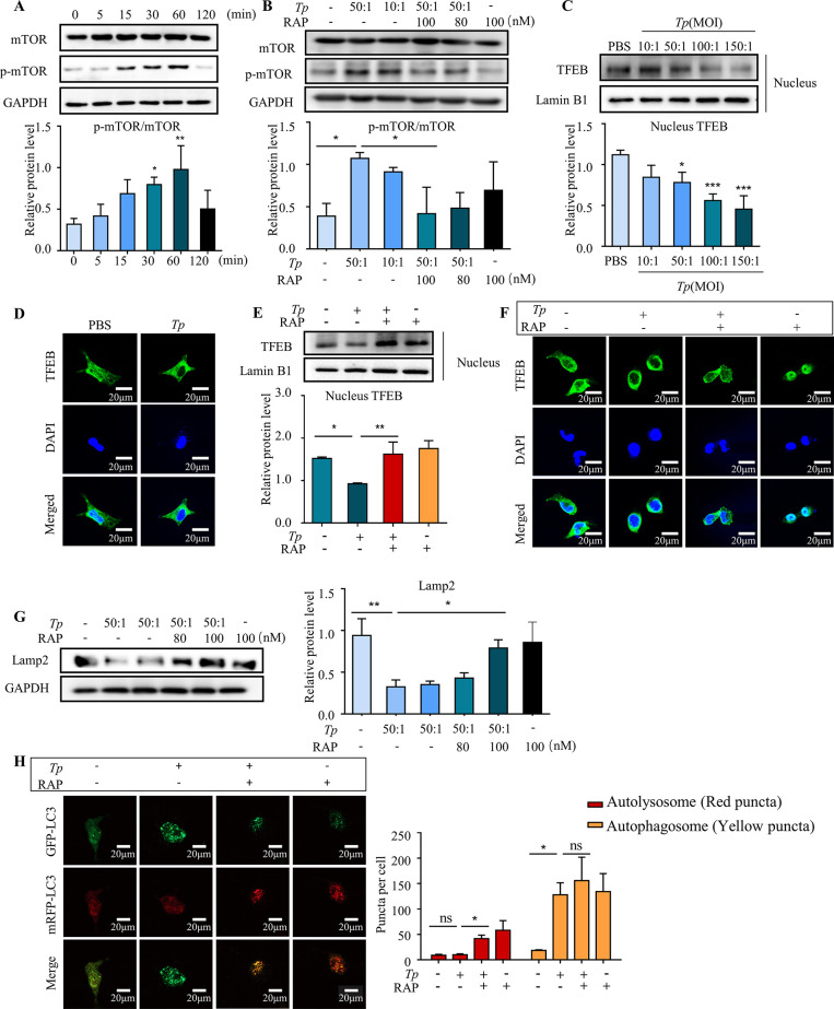
Fig 5. Tp blocks the autophagic flux in HMC3 cells by inhibiting TFEB nuclear translocation and reducing lysosomal biosynthesis via activation of the mTORC1 signalling pathway.
To assess mTOR activation in Tp-treated HMC3 cells, HMC3 cells were cultured with Tp (MOI of 50:1) for different times, and Western blotting was utilized to analyse mTOR signalling pathway components. To investigate the effect of mTORC1 signalling on lysosome biosynthesis and TFEB transcription activity, Western blotting and immunostaining were utilized to measure the expression of lysosome-associated membrane glycoprotein 2 (Lamp2) and nuclear levels of TFEB. To investigate the effect of mTORC1 signalling on the autophagic flux, a tandem fluorescent-labelled plasmid pmCherry-EGFP-LC3B construct was transfected into HMC3 cells. Then, HMC3 cells were pretreated with RAP (rapamycin, mTORC1 inhibitor, 100 nM) for 2 h and cocultured with Tp (MOI of 50:1) for 24 h. Finally, the results were observed under a confocal microscope. (A) Total mTOR protein levels and phosphorylated mTOR protein levels after HMC3 cells were treated with Tp (MOI of 50:1) for different times were measured by Western blotting. (B) HMC3 cells were pretreated with the mTORC1 signalling inhibitor RAP (rapamycin, 100 nM) for 2 h and then cocultured with Tp (MOI of 50:1) for 1 h. The effects of RAP on mTOR signalling were measured by Western blotting. (C) The nuclear levels of TFEB after HMC3 cells were treated with different concentrations of Tp for 24 h was measured by Western blotting. (D) HMC3 cells were treated with Tp (MOI of 50:1) for 24 h and immunostained for TFEB. Scale bar = 100 μm. (E) HMC3 cells were pretreated with the mTORC1 signalling inhibitor RAP (rapamycin, 100 nM) for 2 h and then cocultured with Tp (MOI of 50:1) for 24 h. The effects of RAP on the nuclear levels of TFEB were measured by Western blotting. (F) HMC3 cells were pretreated with the mTORC1 signalling inhibitor RAP (rapamycin, 100 nM) for 2 h, cocultured with Tp (MOI of 50:1) for 24 h, and immunostained for TFEB. Scale bar = 20 μm. (G) HMC3 cells were pretreated with the mTORC1 signalling inhibitor RAP (rapamycin, 100 nM) for 2 h and then cocultured with Tp (MOI of 50:1) for 24 h. The effects of RAP on Lamp2 expression were measured by Western blotting. (H) HMC3 cells were transfected with the pmCherry-EGFP-LC3B plasmid, pretreated with the mTORC1 signalling inhibitor RAP (rapamycin, 100 nM) for 2 h and cocultured with Tp (MOI of 50:1) for 24 h. The effects of RAP on the changes in the autophagic flux in HMC3 cells were analysed by confocal fluorescence microscopy. Scale bar: 20 μm. The data are presented as the mean ± SD of three independent experiments. *P < 0.05, **P < 0.01, ***P < 0.001.

