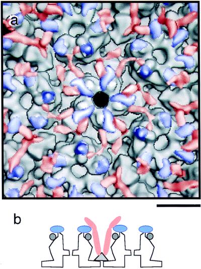
FIG. 8.
(a) Region surrounding the fivefold axis showing the interactions of the two major tegument proteins with the SCMV capsid. The capsomer-capping tegument protein is shown as blue, and the triplex-associated tegument protein is shown as red. The segmentation was achieved by removing noise from the two difference maps (Fig. 5c and d) and then recombining them with the nuclear B-capsid image. Bar, 10 nm. (b) Schematic summary of the interactions that link the tegument to the capsid. The capsomer-capping tegument protein (blue) binds to the major capsid protein on top of the capsomer protrusions. Two copies of the triplex-binding tegument protein (red) diverge from each triplex (the triplex is shown in panel b as a triangle). This elongated protein contacts the capsomer-capping protein (blue) on two different capsomers and then extend outwards. This protein may have an additional domain or domains that extend beyond the portions sketched in panel b, which are not well visualized in our density map (panels b and c) on account of flexibility or otherwise poor ordering.

