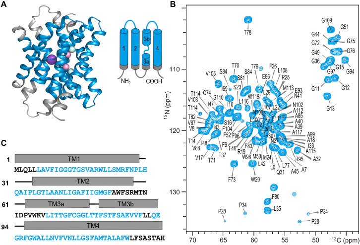
Fig. 1. High-resolution ssNMR spectrum of Fluc-Ec1.
(A) Structural model of Fluc-Ec1 based on the crystal structure of the Fluc channel (PDB ID: 5NKQ). Fluoride ions are shown as pink (F1 sites) or cyan (F2 sites) spheres, and sodium as a purple sphere. (B) Assigned 2D NiCAi correlation spectrum of Fluc-Ec1 in the presence of 150 mM F−. (C) Amino acid sequence of Fluc-Ec1 with transmembrane (rectangle) and loop (short line) annotation based on the crystal structure indicated above the sequence. Assigned residues are highlighted in blue, and unassigned residues are shown in black. ppm, parts per million.

