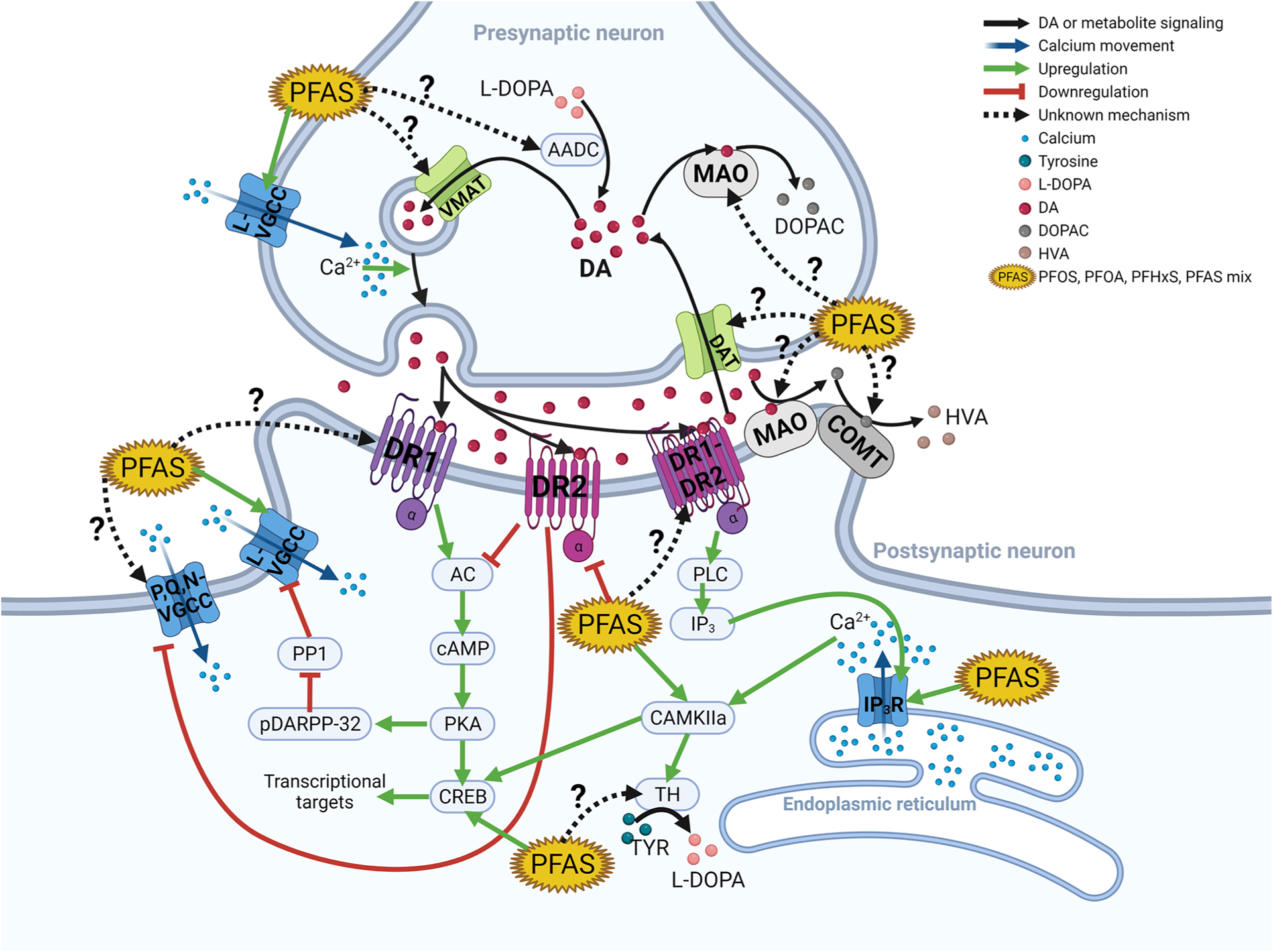
Figure 1.

PFAS neurotoxicity through dopamine and calcium signaling. This figure illustrates the synaptic pathways connecting PFAS, dopamine, and calcium. The key concept is that dopamine, like glutamate, is a target for PFAS-induced modulation of calcium currents. Importantly, PFAS interacts with many known proteins that are a part of dopaminergic postsynaptic signal transduction, but future studies need to investigate the interaction of PFAS and dopamine receptors (DRs) 1 and 2 and the DR1–2 dimer as well as DR3, −4, and −5 (not depicted in the figure). Some evidence suggests DR2 activity, protein levels, and RNA expression may decrease with PFAS exposure. This would result in PFAS inhibiting DR2’s inhibition of AC, resulting in the upregulation of the L-VGCC’s activity. PFAS has been extensively demonstrated to induce increased cytosolic calcium through the L-VGCC and to a lesser extent in the IP3R. Future studies also need to investigate PFAS’s effect on the monoamine metabolic enzymes and transporters. AADC, aromatic amino acid decarboxylase; AC, adenylyl cyclase; CAM, calmodulin; CAMKIIa, CAM kinase IIa; cAMP, cyclic adenosine monophosphate; COMT, catechol-O-methyltransferase; CREB, cAMP-response element-binding; DA, dopamine; DAT, dopamine transporter; DOPAC, 3,4-dihydroxyphenylacetic acid; DR1, dopamine receptor 1; DR1-DR2, DR1–DR2 dimer; DR2, dopamine receptor 2; IP3, inositol triphosphate; IP3R, inositol triphosphate receptor; L-DOPA, l-3,4-dihydroxyphenylalanine; HVA, homovanillic acid; L-VGCC, L-type voltage-gated calcium channel; MAO, monoamine oxidase; pDARPP-32, dopamine and cAMP-regulated phosphor-protein 32 kDa; PKA, protein kinase A; PLC, protein lipase C; P,Q,N-VGCC, P,Q,N-type voltage-gated calcium channel; PP1, protein phosphatase 1; VMAT, vesicular monoamine transporter.
