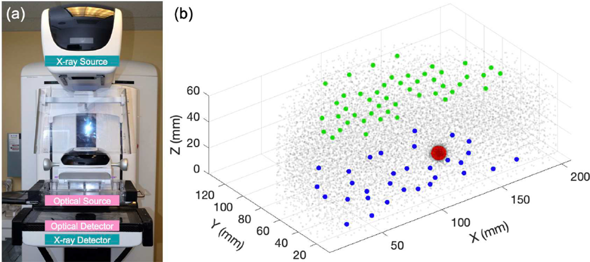
Fig. 1.

(a) Photo of the DOT system integrated with a clinical digital breast tomosynthesis (DBT) device in the transmission geometry, i.e., the DOT source and detector probes arranged on the opposite sides of the compressed breast. (b) A sample phantom geometry with an 8-mm diameter inclusion embedded within a breast-shaped boundary represented by cloud points of the dual-resolution mesh nodes, magenta dots in the fine inclusion region and pale gray dots in the rest of the geometry, respectively. 48 CW sources (green dots) and 32 detectors (blue dots) are plotted as overlays on the top and bottom of the phantom, i.e., superior and inferior in the patient position orientation, respectively.
