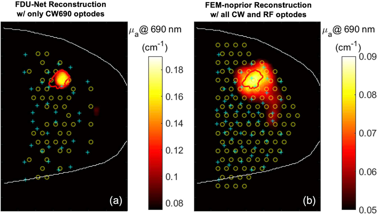
Fig. 9.

Recovered optical images of a patient with a malignant tumor using (left) FDU-Net model trained entirely on simulated data and (right) FEM-based conventional method. White line – breast contour; Red line – tumor marking spatially transformed from ROI drawn by a breast radiologist on separately acquired clinical x-ray mammogram; Yellow circle – source optode; Cyan cross – detector optode. Note that more DOT measurement data were used in the FEM-based reconstruction.
