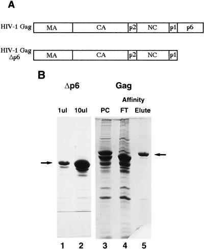
FIG. 1.
Schematic diagrams of proteins expressed in E. coli and purified proteins. (A) Schematic diagram of HIV-1 Gag and Gag Δp6 used for in vitro assembly. (B) Coomassie blue-stained SDS-polyacrylamide gel of purified proteins. Lanes: 1 and 2, 10 and 100 μg of purified HIV-1 Gag Δp6, respectively; 3, HIV-1 Gag purified in the same manner as Gag Δp6; 4 and 5, anti-p6 antibody affinity purification of HIV-1 Gag; 4, flow-through (FT) from the affinity column; 5, affinity-purified HIV-1 Gag (eluted with p6 peptide). Arrows indicate the protein band referred to in each lane. PC, purified on a phosphocellulose cation-exchange column.

