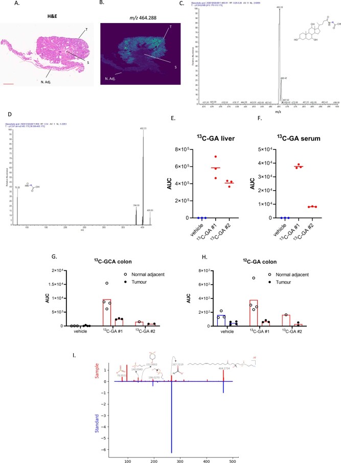
Extended Data Fig. 3. Tumour epithelial specific accumulation of Lyso-PE 17:1.
(A) H&E of distal colon tissue with an Apc-deficient tumour [same H&E as shown in Fig. 2B and Extended Data Fig. 2A; N. Adj.: normal adjacent tissue; S: stroma; T: tumour tissue]. Tissues analysed for n = 4 animals; n = 1 shown. Scale bar: 1 mm. (B) specific accumulation of an ion with m/z 464.288 in the tumour epithelial compartment of APC tumours as detected by MALDI-MSI. The two top tentative parent ion identifications (that is glycocholic acid and LysoPE 17:1) obtained from a publicly available database (https://hmdb.ca) were experimentally validated. (C) A 13C-glycocholic acid (13C-GA) standard solution was infused to detect the parent ion (blue dot indicates 13C label) using Triple Quadrupole Mass Spectrometry, and (D) a Multiple Reaction Monitoring (MRM) method was developed to study the abundance of 13C-GA in tissues via its a diagnostic ion of 75 Da (blue dot indicates 13C label). Tumour-bearing APC mice were administered 13C-GA (75 mg/kg p.o.; n = 2) or vehicle (HPMC/Tween:DMSO; v-v 90:10; n = 1). After 8.5 hours, serum, liver and distal colon were harvested and processed for LC-MS. 13C-GA was detected in (E) liver, (F) serum, (G) normal adjacent colonic tissue and tumour tissues. No increased abundance of (G) 13C-GA or (H) 12C-GA was observed in tumour tissues compared with normal adjacent colonic tissue. Panels E-H show the mean of metabolic extractions of the multiple tissue fragments for each mouse, each dot represents data obtained from a single tissue fragment, or serum extract. Liquid extraction surface analysis tandem mass spectrometry was applied to study fragmentation of the m/z 464.288 precursor ion, which showed matched ions to the predicted fragmentation pattern of LysoPE 17:1, as well as to the (I) fragmentation of a commercially available standard further supporting the ID of this metabolite of interest. All highlighted m/z of fragments and precursor ions on the mass spectra are present in both standard and sample.

