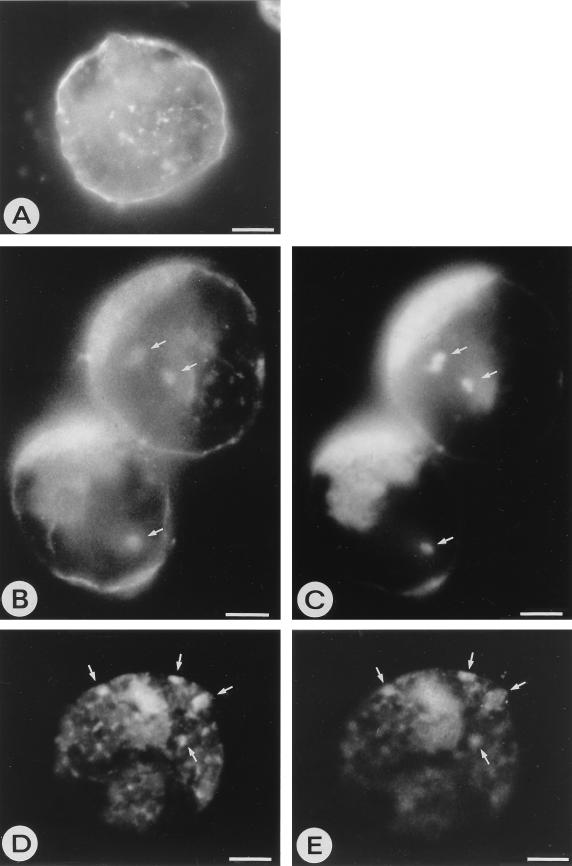
FIG. 5.
Immunofluorescence images of N. rustica protoplasts. (A) Healthy protoplast labeled with JIM84 antiserum against the plant Golgi system, showing individual Golgi stacks as small clusters throughout the cytoplasm. The plasma membrane is also labeled with JIM84 due to transport of the epitope-containing Golgi proteins. (B and D) TSWV-infected protoplasts at 30 h p.i., labeled with JIM84 antiserum. (C and E) The same infected protoplasts labeled with mixed antisera against G1 and G2. Areas of clear colocalization of the viral glycoproteins with the Golgi system are indicated with arrows. Cloudy areas within cells represent the autofluorescence background. Bars, 5 μm.

