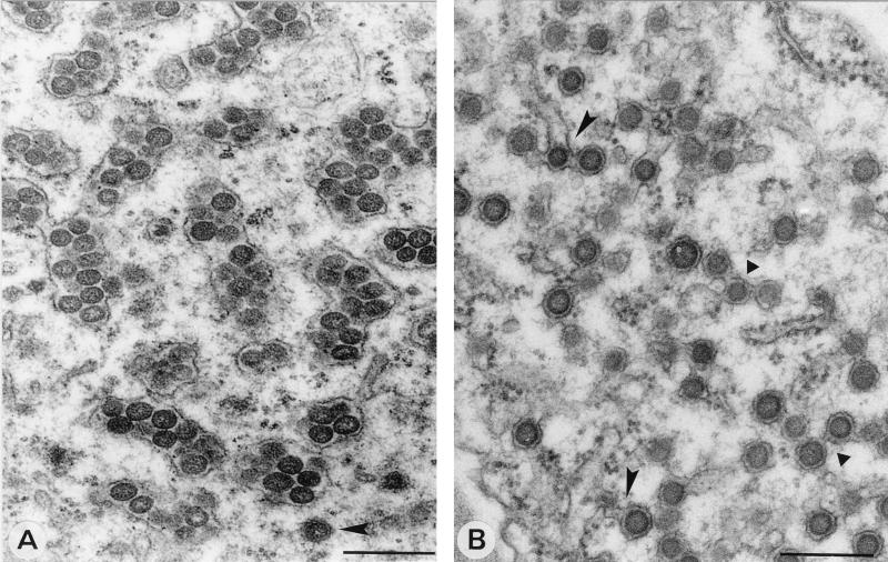
FIG. 8.
Formation of SEV by fusion of DEV. (A) Clustered SEV inside smooth and rough membranes, as found in late stages of TSWV infections of N. rustica protoplasts at 40 h p.i. Note the DEV at the bottom of the image (arrowhead). (B) Image from a TSWV local lesion in petunia showing DEV particles fusing with each other (triangles) and with ER membranes identified by ribosomes on the surface (arrowheads). Bars, 200 nm.

