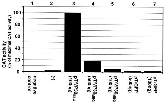
FIG. 6.
Exchange of EBOV VP30 by MBGV VP30. HeLa cells were infected with MVA-T7 and transfected with DNA and RNA as described under Fig. 2. At 2 days p.i., the cell lysates were analyzed for CAT activity. DNA transfection was performed with the following plasmids: 500 ng of pT/NPEBO, 500 ng of pT/VP35EBO, and 1 μg of pT/LEBO; pT/VP30MBG and pT/GFP were added as indicated in the figure. As a negative control, pT/LEBO was omitted (negative control). For RNA transfection, minigenome 3E-5E was used. For the CAT assay, 1.5 μl of each cell lysate (except lane 3) was used. For the sample shown in lane 3, only a fifth of this amount was subjected to CAT assay. After quantification, the obtained value for the sample shown in lane 3 was multiplied by five and set as the maximal CAT activity (100%). −, DNA transfection with pT/NPEBO, pT/VP35EBO, and pT/LEBO.

