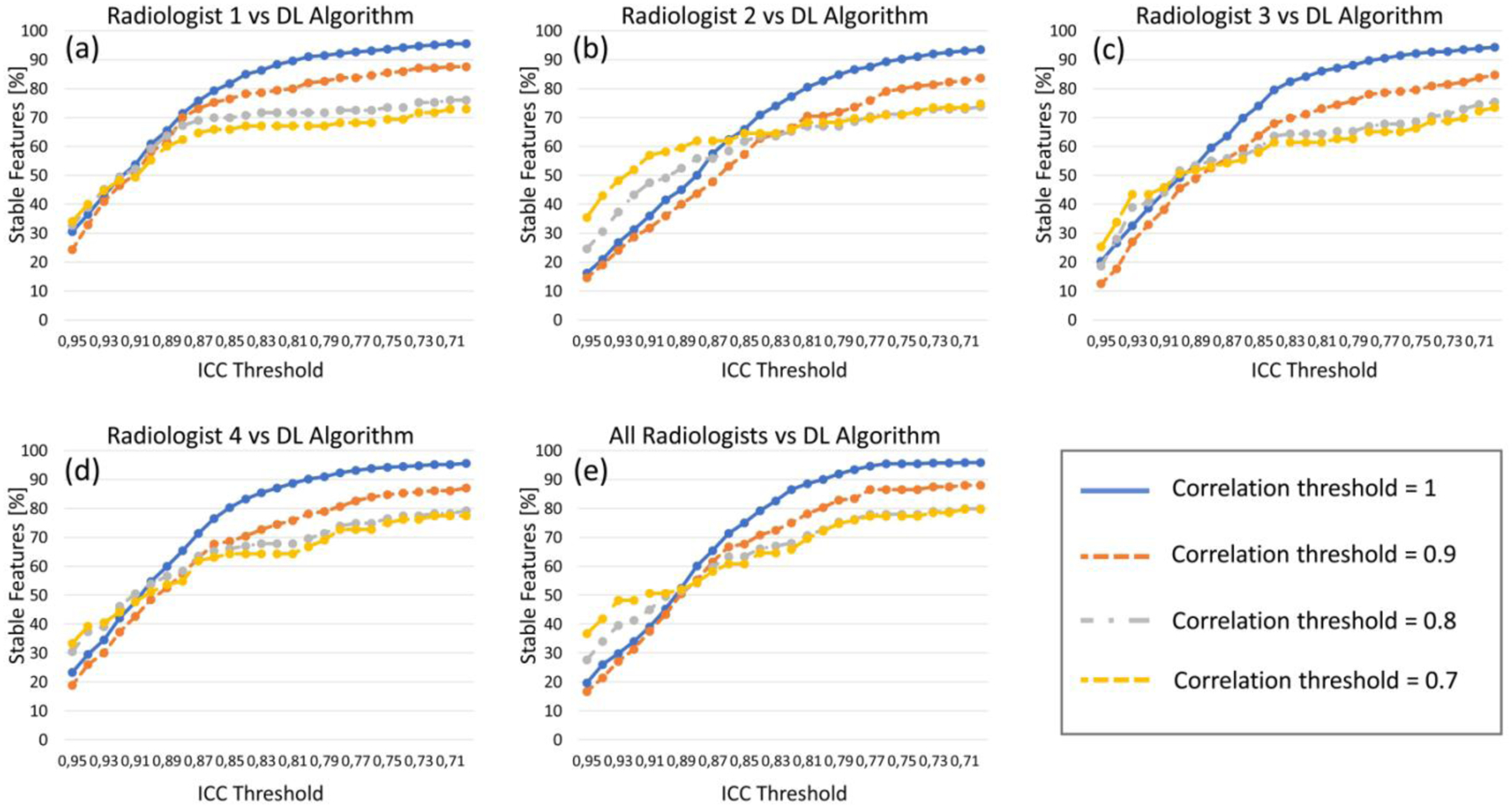
Fig. 6.

Results of radiomic feature stability analysis. Each graph shows the percentage of features (y axis) having different ICC values (x axis), after eliminating the highly correlated features for four thresholds of correlation (1, 0.9, 0.8, 0.7). (a)-(d) show the feature stability for the four radiologists’ annotations (each compared with the DL algorithm), while (e) shows the stability for the deep learning-based segmentation compared to all radiologists together.
