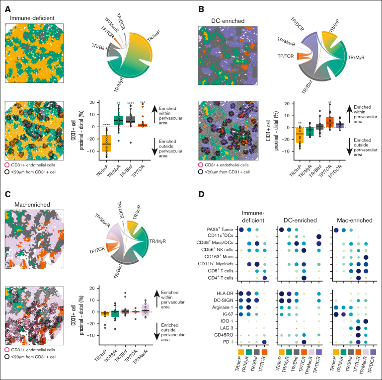
Figure 3.
Cases with immune-deficient, DC-enriched, and Mac-enriched aggregate TIMEs have distinct topographical structures and express distinct functional markers. (A) (Top left) Representative CNT map of a DLBCL with an immune-deficient TIME and colored per the CNT assignment of the cells as TR/ImP (yellow), TR/MyR (green), or TR/BInf (gray). (Top right) Chord diagram integrating all contacts between cells assigned to different CNTs for all cases with an immune-deficient TIME. Broader bands reflect greater number of contacts between cells. (Bottom left) Representative CNT map of a DLBCL with an immune-deficient TIME with an overlay of CD31+ endothelial cells (red outlines) and cells within a 20 μm radial distance from CD31+ cells (black outlines). (Bottom right) The differences in the percentages of cells assigned to the indicated CNT within (proximal) or outside (distal) a 20 μm radial distance from a CD31+ cell. Values for individual cases (dots), median values (horizontal line), and 25th and 75th centiles (boxes) are indicated. Positive values represent enrichment within the perivascular region, whereas negative values represent enrichment outside the perivascular area. Only significant P-values are annotated ∗P < .05; ∗∗P < .01; ∗∗∗P < .001; ∗∗∗∗P < .0001 using the paired 2-sided Wilcoxon sign-rank test. (B) (Top left) Representative CNT map from a DLBCL with a DC-enriched TIME and colored per the CNT assignment of the cells as TP/DCR (blue), TP/TCR (orange), TR/BInf (gray), TR/MyR (green), or TR/ImP (yellow). (Top right) Chord diagram integrating all contacts among cells in neighborhoods assigned to different CNTs for all cases with a DC-enriched TIME. Broader bands reflect greater numbers of contacts between cells. (Bottom left) Representative CNT map from a DLBCL with a DC-enriched TIME with an overlay of CD31+ endothelial cells (red outlines) and cells within a 20 μm radial distance from CD31+ cells (black outlines). (Bottom right) The differences in the percentages of cells assigned to the indicated CNT within (proximal) or outside (distal) a 20 μm radial distance from a CD31+ cell. Values for individual cases (dots), median values (horizontal line), and 25th and 75th centiles (boxes) are indicated. Positive values represent enrichment within the perivascular region, while negative values represent enrichment outside the perivascular area. Only significant P-values are annotated. ∗∗P < .01, using the paired 2-sided Wilcoxon sign-rank test. (C) (Top right) Representative CNT map of a DLBCL with a Mac-enriched TIME that is color-coded per the CNT assignment of cells as TP/MacR (lavender), TP/TCR (orange), TR/ImP (yellow), TR/MyR (green), or TR/BInf (gray). (Top right) Chord diagram integrating all contacts between cells assigned to different CNTs for all cases with a Mac-enriched TIME. Broader bands reflect greater number of contacts between cells. (Bottom left) Representative CNT map of a DLBCL with a Mac-enriched TIME with an overlay of CD31+ endothelial cells (red outlines) and cells within a 20 μm radial distance from a CD31+ cell (black outlines). (Bottom left) The differences in the percentages of cells assigned to the indicated CNT within (proximal) or outside (distal) a 20 μm CD31+ cell. Values for individual cases (dots), median values (horizontal line), and 25th and 75th centiles (boxes) are indicated. Positive values represent enrichment within the perivascular region, while negative values represent enrichment outside the perivascular region. No P-values were significant using the paired 2-sided Wilcoxon sign-rank test. (D) The relative cell densities (in cells per mm2) of each indicated cell lineage (top) and the relative expression (in ion counts) of each indicated functional biomarker (bottom) for each indicated CNT (columns) and separated based on the aggregate TIME category (immune-deficient, DC-enriched, and Mac-enriched). Higher cell density (top) and ion counts (bottom) are indicated by larger circle size and darker color.

